Review
. 2013 Apr;24(4):258-66.
doi: 10.1016/j.semcdb.2012.12.005. Epub 2013 Jan 3.

Under lock and key: spatiotemporal regulation of WASP family proteins coordinates separate dynamic cellular processes

Affiliations
Review

Under lock and key: spatiotemporal regulation of WASP family proteins coordinates separate dynamic cellular processes

Lauren E Burianek et al. Semin Cell Dev Biol. 2013 Apr.

Abstract

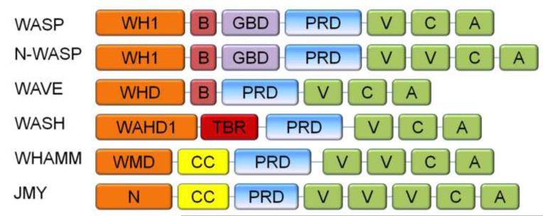
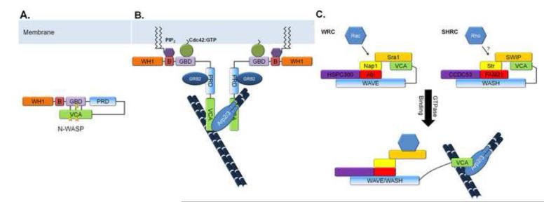
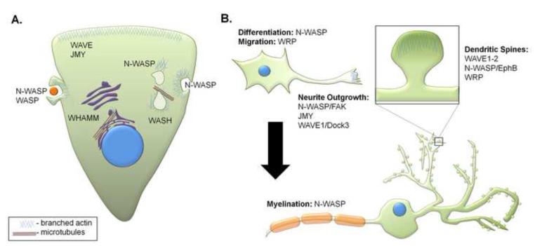
WASP family proteins are nucleation promoting factors that bind to and activate the Arp2/3 complex in order to stimulate nucleation of branched actin filaments. The WASP family consists of WASP, N-WASP, WAVE1-3, WASH, and the novel family members WHAMM and JMY. Each of the family members contains a C-terminus responsible for their nucleation promoting activity and unique N-termini that allow for them to be regulated in a spatiotemporal manner. Upon activation they reorganize the cytoskeleton for different cellular functions depending on their subcellular localization and regulatory protein interactions. Emerging evidence indicates that WASH, WHAMM, and JMY have functions that require the coordination of both actin polymerization and microtubule dynamics. Here, we review the mechanisms of regulation for each family member and their associated in vivo functions including cell migration, vesicle trafficking, and neuronal development.


Figures

Fig. 1
Fig. 1. Domain structures of WASP family proteins
All of the WASP family members exhibit a proline rich region and a VCA tail in the C-terminus, but contain unique N-termini. WH1: WASP Homology domain, B: Basic domain, GBD: GTPase binding domain, PRD: Proline rich domain, V: Verprolin homology, C: Connecting sequence, A: Acidic sequence, WHD: WAVE homology domain, WAHD1: WASH Homology domain, TBR: Tubulin binding region, WMD: WHAMM membrane-interacting domain, N: N-terminal region
Fig. 2
Fig. 2. Regulation of WASP family proteins
(a) WASP and N-WASP are autoinhibited through intramolecular interactions between the GBD and VCA domain. (b) This autoinhibition can be released upon Cdc42 or PIP2 binding, or upon phosphorylation (phosphorylation sites marked with stars). Dimerization of VCA domains and interactions with the Arp2/3 complex leads to robust activation of branched actin nucleation. WASP can also self-dimerize in an inhibitory manner (not shown for clarity). GRB2 binds to the PRD and stabilizes the active WASP proteins. (c) WAVE and WASH proteins are sequestered in regulatory complexes, termed WRC and SHRC respectively. The VCA domain of WAVE is sequestered in a complex with Sra1. Rac1 binds to Sra1 to induce a conformational change that releases the VCA domain to allow for Arp2/3 activation. The SHRC makes up a structurally related inhibitory complex that is thought to behave in a similar manner.
Fig. 3
Fig. 3. Cellular roles of WASP family proteins
(a) WASP family proteins coordinate membrane and cytoskeletal events, such as migration, endocytosis, and vesicle transport. WAVE and JMY play roles in cellular motility at the leading edge. WASP and N-WASP are involved in phagocytosis and endocytosis. N-WASP also leads to endosomal “rocketing”. WASH is responsible for pinching recycling vesicles off of endosomes, and WHAMM is essential for proper anterograde transport from the ER to the Golgi; both of these processes require interactions with microtubules. (b) WASP family proteins are involved in several aspects of neuronal development, including differentiation, migration, neurite outgrowth and myelination. In mature neurons, WASP and WAVE proteins are involved in the development and morphology of dendritic spines.

Similar articles

Cited by

References

    1. Fowler WE, Aebi U. A consistent picture of the actin filament related to the orientation of the actin molecule. The Journal of cell biology. 1983;97:264–9. - PMC - PubMed
    1. Mullins RD, Stafford WF, Pollard TD. Structure, subunit topology, and actin-binding activity of the Arp2/3 complex from Acanthamoeba. The Journal of cell biology. 1997;136:331–43. - PMC - PubMed
    1. Mullins RD, Heuser JA, Pollard TD. The interaction of Arp2/3 complex with actin: nucleation, high affinity pointed end capping, and formation of branching networks of filaments. Proceedings of the National Academy of Sciences of the United States of America. 1998;95:6181–6. - PMC - PubMed
    1. Svitkina TM, Verkhovsky AB, McQuade KM, Borisy GG. Analysis of the actin-myosin II system in fish epidermal keratocytes: mechanism of cell body translocation. The Journal of cell biology. 1997;139:397–415. - PMC - PubMed
    1. Korobova F, Svitkina T. Molecular architecture of synaptic actin cytoskeleton in hippocampal neurons reveals a mechanism of dendritic spine morphogenesis. Molecular biology of the cell. 2010;21:165–76. - PMC - PubMed

Publication types

MeSH terms

Substances