Review
. 2013 Feb;62(1):69-79.
doi: 10.1093/jmicro/dfs097. Epub 2013 Jan 4.

Confessions of an icosahedral virus crystallographer

Affiliations
Review

Confessions of an icosahedral virus crystallographer

John E Johnson. Microscopy (Oxf). 2013 Feb.

Abstract

This is a personal history of my structural studies of icosahedral viruses that evolved from crystallographic studies, to hybrid methods with electron cryo-microscopy and image reconstruction (cryoEM) and then developed further by incorporating a variety of physical methods to augment the high resolution crystallographic studies. It is not meant to be comprehensive, even for my own work, but hopefully provides some perspective on the growth of our understanding of these remarkable biologic assemblies. The goal is to provide a historical perspective for those new to the field and to emphasize the limitations of any one method, even those that provide atomic resolution information about viruses.


Figures
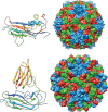
Fig. 1.
Fig. 1.
Comparison of the subunit tertiary structure (ramped in color from blue at the N-terminus to red at the C-terminus) and T = 3 particle quaternary structure of SBMV (top) and TBSV. Although there was no detectable sequence homology, the structures of the shell-forming domain were virtually superimposable. TBSV has an additional domain at the C-terminus of the subunit that adds the surface protrusions to the particle. The quaternary structures of the contiguous shell domains in SBMV and TBSV are also nearly identical. The maximum dimension of the SBMV particle is approximately 300 Å.
Fig. 2.
Fig. 2.
Surface-shaded representations of the 23 Å cryoEM reconstructions of native CPMV and CPMV with a monoclonal antibody bound to it. The known crystal structure of CPMV allowed the identification of the ‘footprint’ of the antibody on to the surface residues of the virus [31].
Fig. 3.
Fig. 3.
The structure of Pariocota virus, a typical Nodavirus. At left is the fold of the A subunit (blue subunit in the quaternary structure and clustered about the 5-fold symmetry axes of the icosahedron). The structure is ramped in color as in Fig. 1. The T = 3 quaternary structure has characteristic protrusions at the quasi 3-fold symmetry axes (middle). At right is a cutaway of the capsid showing the ordered duplex RNA and those portions of the subunit polypeptide that interact with the RNA. The gamma peptides are in blue and can be seen to lie in cavities within the protein shell, shown as density from a cryoEM reconstruction.
Fig. 4.
Fig. 4.
The subunit tertiary structure (color ramped as in Fig. 1) for Nudaurelia Omega Capensis virus is shown at left. The transition from procapsid to capsid for the T = 4 particle is depicted on the right. The color of the quaternary structure is ramped by radius from red to blue. The procapsid particle is about 490 Å in diameter with the subunits clustered as dimmers. After maturation at pH 5.0, the particle size decreases to 410 Å, and the subunits are clustered as trimers. An autocatalytic cleavage is activated when the particles compact [37].
Fig. 5.
Fig. 5.
The HK97 virus-like particle assembly and maturation pathway that is followed, when only the coat protein and protease are co-expressed in Escherichia coli. At the top are all the intermediates that have been characterized by crystallography and/or cryoEM. Below is shown the processing that occurs to the capsid protein, gp5, and the residues that form the autocatalytic crosslink. At right is an enlargement of the final mature particle indicating that the cross-linked gp5 proteins mechanically chain link the particle together. Each ring of the same color corresponds to five or six subunits chain linked together by the isopeptide bond formed by the side chains of Asn 356 and Lys169.

Similar articles

Cited by

References

    1. Harrison S C. A point-focusing camera for single-crystal diffraction. J. Appl. Cryst. 1968;1:84–90. doi:10.1107/S0021889868005054. - DOI
    1. Harrison S C. Structure of tomato bushy stunt virus. I. The spherically averaged electron density. J. Mol. Biol. 1969;42:457–483. doi:10.1016/0022-2836(69)90236-8. - DOI - PubMed
    1. Harrison S C. Structure of tomato bushy stunt virus at 25 Å resolution. Cold Spring Harb. Symp. Quant. Biol. 1971;36:495–501. doi:10.1101/SQB.1972.036.01.063. - DOI - PubMed
    1. Harrison S C, Jack A. Structure of tomato bushy stunt virus. Three-dimensional x-ray diffraction analysis at 16 A resolution. J. Mol. Biol. 1975;97:173–191. doi:10.1016/S0022-2836(75)80033-7. - DOI - PubMed
    1. Jack A, Harrison S C, Crowther R A. Structure of tomato bushy stunt virus. II. Comparison of results obtained by electron microscopy and x-ray diffraction. J. Mol. Biol. 1975;97:163–172. doi:10.1016/S0022-2836(75)80032-5. - DOI - PubMed

MeSH terms

LinkOut - more resources