

Incidental renocolic fistula with xanthogranulomatous pyelonephritis
- PMID: 23291328
- PMCID: PMC3540218
- DOI: 10.1016/j.ijscr.2012.08.003
Incidental renocolic fistula with xanthogranulomatous pyelonephritis
Abstract
Introduction: We report the case of a 66-year-old female undergoing elective nephrectomy for a non-functioning kidney in whom an incidental renocolic fistula was detected.
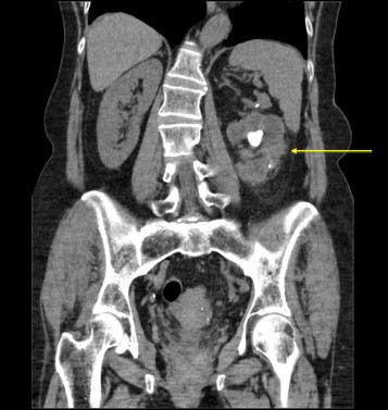
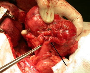
Presentation of case: She presented with recurrent urinary tract infections and left flank pain. Investigations revealed a nonfunctioning left kidney with a large staghorn calculus and features suggestive of xanthogranulomatous pyelonephritis (XPG). At nephrectomy, an incidental renocolic fistula was found and excised.
Discussion: XGP is a rare, chronic inflammatory disorder of the kidney characterized by a destructive mass invading the renal parenchyma. Renocolic fistulae complicating XGP are uncommon and not widely reported in the literature.
Conclusion: Herein, we describe a case of XGP with renocolic fistula formation, its management and a review of the literature.
Copyright © 2012 Surgical Associates Ltd. Published by Elsevier Ltd. All rights reserved.
Figures

References
-
- Schlagenhaufer F. Ueber eigentumliche Staphylomykosen der Nieren und des pararenalen Bindegewebes. Frankfurter Zeitschrift fur Pathologie. 1916;19:139–148.
-
- Yazaki T., Ishikawa S., Ogawa Y., Takahashi S., Nemoto S., Rinsho K. Xanthogranulomatous pyelonephritis in childhood: case report and review of English and Japanese literature. Journal of Urology. 1982;127:80–83. - PubMed
-
- Parsons M.A., Harris S.C., Grainger R.G., Ross B., Smith J.A., Williams J.L. Fistula and sinus formation in xanthogranulomatous pyelonephritis. A clinicopathological review and report of four cases. British Journal of Urology. 1986;58(5):488. - PubMed
-
- Zafranloo S., Gerard P.S., Bryk D. Xanthogranulomatous pyelonephritis in children: analysis by diagnostic modalities. Urologic Radiology. 1990;12:18–21. - PubMed
-
- Rafal R.B., Kosovsky P.A., Markisz J.A. Xanthogranulomatous pyelonephritis in an infant. Urology. 1991;37:553–556. - PubMed
LinkOut - more resources
Full Text Sources
Other Literature Sources
Research Materials
