

Natural consequence of post-intervention stent malapposition, thrombus, tissue prolapse, and dissection assessed by optical coherence tomography at mid-term follow-up
- PMID: 23291393
- PMCID: PMC3738096
- DOI: 10.1093/ehjci/jes299
Natural consequence of post-intervention stent malapposition, thrombus, tissue prolapse, and dissection assessed by optical coherence tomography at mid-term follow-up
Abstract
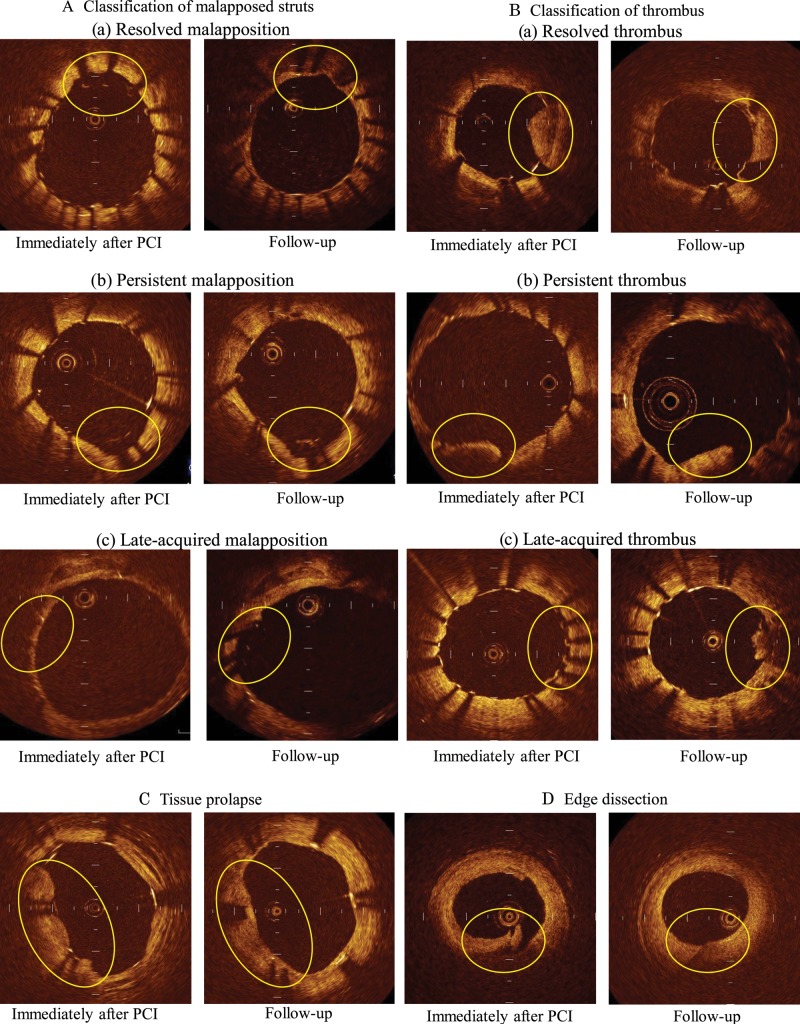
Aims: We performed this study to clarify natural consequences of abnormal structures (stent malapposition, thrombus, tissue prolapse, and stent edge dissection) after percutaneous coronary intervention (PCI).
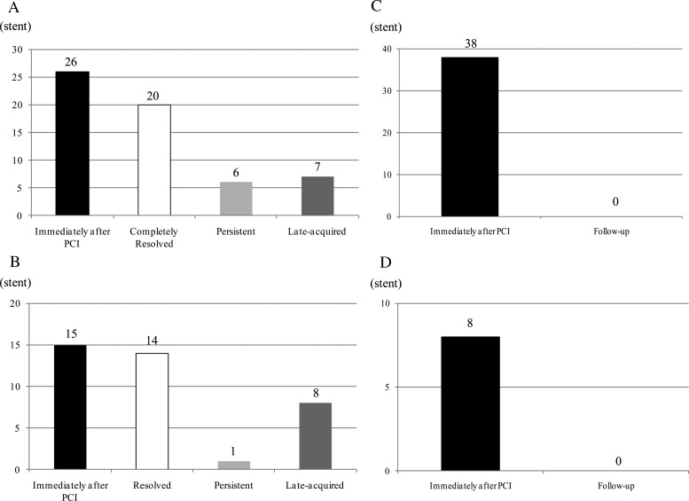
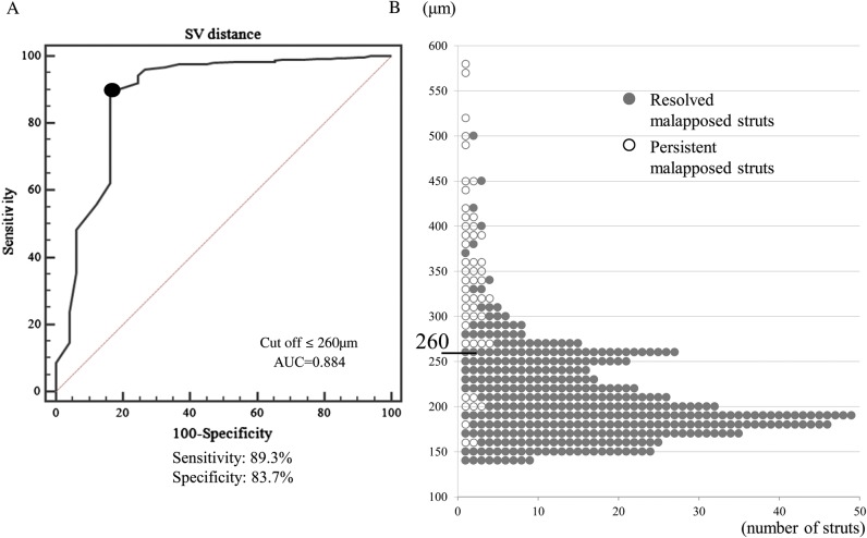
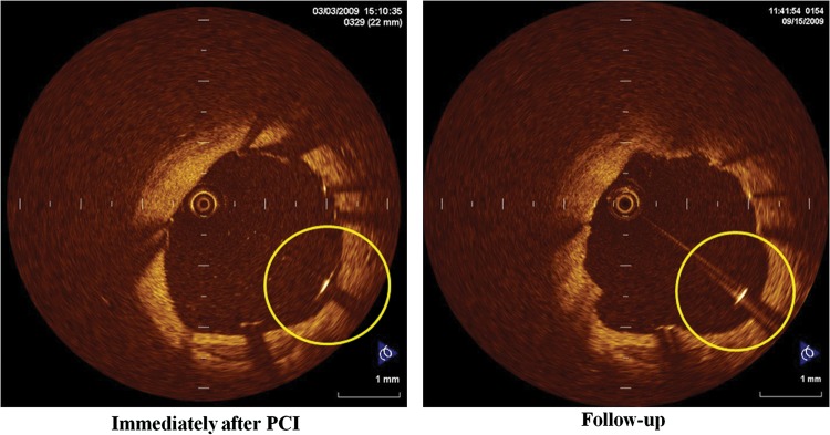
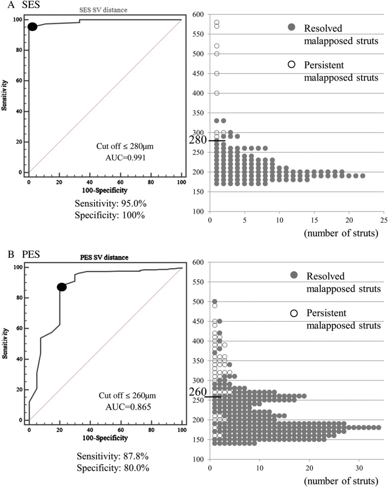
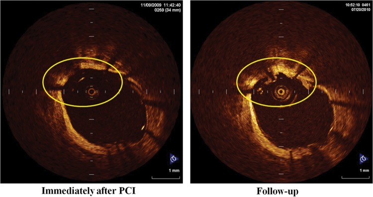
Methods and results: Thirty-five patients treated with 40 drug-eluting stents underwent serial optical coherence tomography (OCT) imaging immediately after PCI and at the 8-month follow-up. Among a total of 73 929 struts in every frame, 431 struts (26 stents) showed malapposition immediately after PCI. Among these, 49 remained malapposed at the follow-up examination. The mean distance between the strut and vessel wall (S-V distance) of persistent malapposed struts on post-stenting OCT images was significantly longer than that of resolved malapposed struts (342 ± 99 vs. 210 ± 49 μm; P <0.01). Based on receiver-operating characteristic curve analysis, an S-V distance ≤260 µm on post-stenting OCT images was the corresponding cut-off point for resolved malapposed struts (sensitivity: 89.3%, specificity: 83.7%, area under the curve = 0.884). Additionally, 108 newly appearing malapposed struts were observed on follow-up OCT, probably due to thrombus dissolution or plaque regression. Thrombus was observed in 15 stents post-PCI. Serial OCT analysis revealed persistent thrombus in 1 stent, resolved thrombus in 14 stents, and late-acquired thrombus in 8 stents. Tissue prolapse observed in 38 stents had disappeared at the follow-up. All eight stent edge dissections were repaired at the follow-up.
Conclusion: Most cases of stent malapposition with a short S-V distance, thrombus, tissue prolapse, or minor stent edge dissection improved during the follow-up. These OCT-detected minor abnormalities may not require additional treatment.
Keywords: Optical coherence tomography; Stent edge dissection; Stent malapposition; Thrombus; Tissue prolapse.
Figures

References
-
- Gonzalo N, Serruys PW, Okamura T, Shen ZJ, Onuma Y, Garcia-Garcia HM, et al. Optical coherence tomography assessment of the acute effects of stent implantation on the vessel wall: a systematic quantitative approach. Heart. 2009;95:1913–9. - PubMed
-
- Kawamori H, Shite J, Shinke T, Otake H, Sawada T, Kato H, et al. The ability of optical coherence tomography to monitor percutaneous coronary intervention: detailed comparison with intravascular ultrasound. J Invasive Cardiol. 2010;22:541–5. - PubMed
-
- Matsumoto D, Shite J, Shinke T, Otake H, Tanino Y, Ogasawara D, et al. Neointimal coverage of sirolimus-eluting stents at 6-month follow-up: evaluated by optical coherence tomography. Eur Heart J. 2007;28:961–7. - PubMed
-
- Guo N, Maehara A, Mintz GS, He Y, Xu K, Wu X, et al. Incidence, mechanisms, predictors, and clinical impact of acute and late stent malapposition after primary intervention in patients with acute myocardial infarction: an intravascular ultrasound substudy of the Harmonizing Outcomes with Revascularization and Stents in Acute Myocardial Infarction (HORIZONS-AMI) trial. Circulation. 2010;122:1077–84. - PubMed
-
- Otake H, Shite J, Ako J, Shinke T, Tanino Y, Ogasawara D, et al. Local determinants of thrombus formation following sirolimus-eluting stent implantation assessed by optical coherence tomography. JACC Cardiovasc Interv. 2009;2:459–66. - PubMed
Publication types
MeSH terms
Substances
LinkOut - more resources
Full Text Sources
Other Literature Sources
Medical
Miscellaneous
