. 2013 Apr;9(4):6095-104.
doi: 10.1016/j.actbio.2012.12.028. Epub 2013 Jan 2.

Primary human chondrocyte extracellular matrix formation and phenotype maintenance using RGD-derivatized PEGDM hydrogels possessing a continuous Young's modulus gradient

Affiliations

Primary human chondrocyte extracellular matrix formation and phenotype maintenance using RGD-derivatized PEGDM hydrogels possessing a continuous Young's modulus gradient

Laura A Smith Callahan et al. Acta Biomater. 2013 Apr.

Abstract

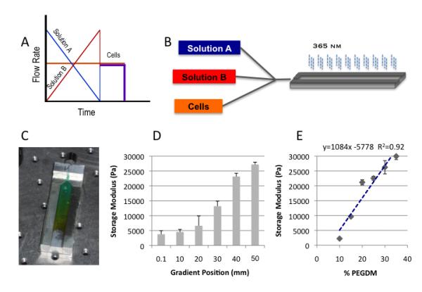
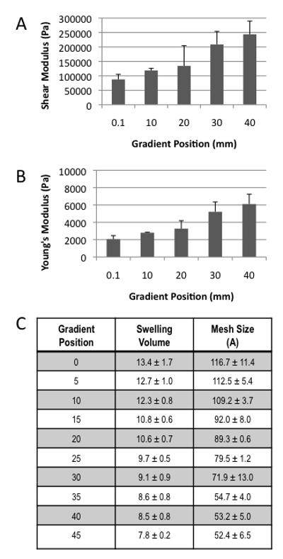
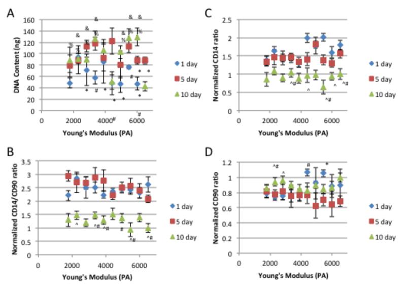
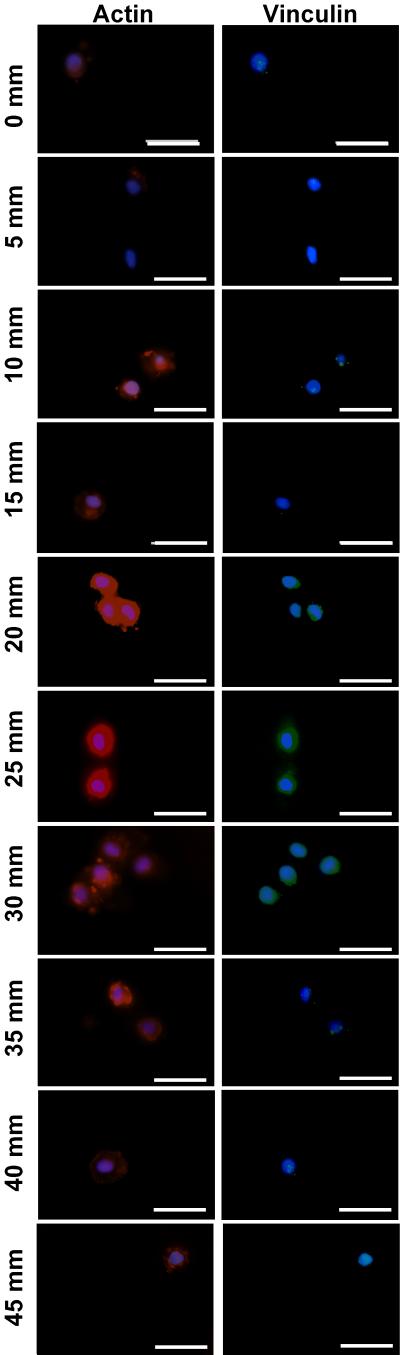
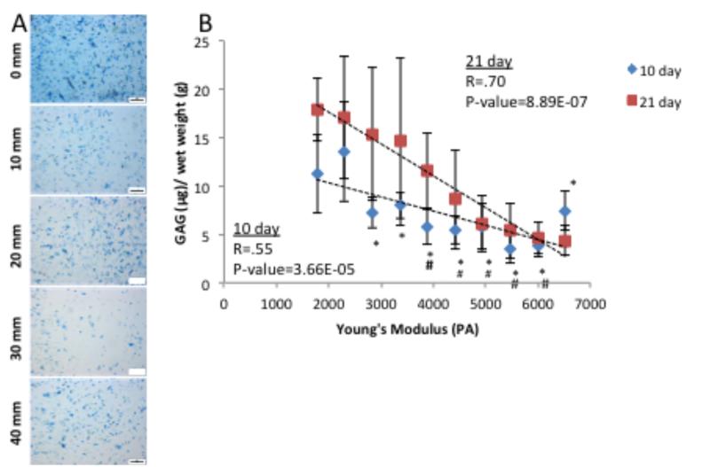
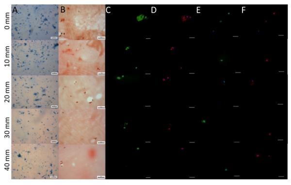
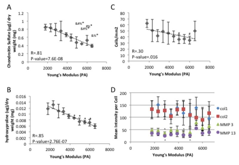
Efficient ex vivo methods for expanding primary human chondrocytes while maintaining the phenotype is critical to advancing the sourcing of autologous cells for tissue engineering applications. While there has been significant research reported in the literature, systematic approaches are necessary to determine and optimize the chemical and mechanical scaffold properties for hyaline cartilage generation using limited cell numbers. Functionalized hydrogels possessing continuous variations in physico-chemical properties are, therefore, an efficient three-dimensional platform for studying several properties simultaneously. Herein we describe a polyethylene glycol dimethacrylate (PEGDM) hydrogel system with a modulus gradient (~27,000-3800 Pa) containing a uniform concentration of arginine-glycine-aspartic acid (RGD) peptide to enhance cell adhesion in order to correlate primary human osteoarthritic chondrocyte proliferation, phenotype maintenance, and extracellular matrix (ECM) production with hydrogel properties. Cell number and chondrogenic phenotype (CD14:CD90 ratios) were found to decline in regions with a higher storage modulus (>13,100 Pa), while regions with a lower storage modulus maintained their cell number and phenotype. Over 3 weeks culture hydrogel regions possessing a lower Young's modulus experienced an increase in ECM content (~200%) compared with regions with a higher storage modulus. Variations in the amount and organization of the cytoskeletal markers actin and vinculin were observed within the modulus gradient, which are indicative of differences in chondrogenic phenotype maintenance and ECM expression. Thus scaffold mechanical properties have a significant impact in modulating human osteoarthritic chondrocyte behavior and tissue formation.


Figures

Figure 1
Figure 1
Depicts the overall framework and assembly of the gradient fabrication system. Schematic of the controlled variation in syringe pump rates coupled with a constant rate of cell infusion which ensures uniform cell density throughout the gradient A); a schematic for the three component (expandable to five) gradient fabrication system B); and an example of the gradient profile that can be generated depicted with a colorimetric gradient for visualization purposes C). The storage modulus can be controlled systematically enabling facile correlations of mechanical properties via position D). The positional values correlate well with values obtained from discrete hydrogel standards of known composition and mass fraction E). Mass fraction, pore size, transitional dimensions are controlled by defined solution mixtures pre-loaded in syringe pumps.
Figure 2
Figure 2
Characterization of all hydrogel physical properties can be correlated to position within the gradient via the highly reproducible fabrication process A) Shear Modulus B) Young’s Modulus and C) Table of swelling volume and calculated mesh size after 24 h of swelling.
Figure 3
Figure 3
Chondrocyte A) DNA content , phenotype maintenance indicator B) CD14/CD90 ratio, C) cellular fraction expressing CD14 and D) cellular fraction expressing CD90 over 10 days of culture. * indicates a p-value ≤ 0.05 compared to the value at day 10 within the same gradient position, # indicates a p-value ≤ 0.05 compared to the value at day 5 within the same gradient position, ^ indicates a p-value ≤ 0.05 compared to the value at day 1 within the same gradient position, % indicates a p-value ≤ 0.05 compared to 6500 Pa Young’s Modulus gradient position at day 10, and & indicates a p-value ≤ 0.05 compared to 4400 Pa Young’s Modulus gradient position at day 10.
Figure 4
Figure 4
Specific staining for the cytoskeletal elements actin (Red), vinculin (Green) and nuclear (blue) after 5 days of culture show distinct morphometric changes as the modulus changes from a low modulus (0 mm) to a high modulus (45 mm) position. Scale bar = 25 μM
Figure 5
Figure 5
Extracellular matrix production by human chondrocytes after 10 days of culture. Images were taken at 10 mm intervals along the length of the modulus gradient. A) Whole mount Alcian blue and B) Sulfated gylcosaminoglycan quantification based on Alcian blue extraction show distinct changes with position in the modulus profile at both 10 day and 21 days. Scale bar = 200 μm. # indicates a p-value ≤ 0.05 compared to 1700 Pa Young’s Modulus gradient position, and * indicates a p-value ≤ 0.05 compared to 2300 Pa Young’s Modulus gradient position
Figure 6
Figure 6
Extracellular matrix production by human chondrocytes after 3 weeks of culture. Images where taken every 5mm down the length of the gradient A) Whole mount Alcian Blue staining. Scale bar = 200 μm. B) Sirius red staining of histological sections. Scale bar = 50 μm. C) Type 2 Collagen (green) and nuclear (blue) immunofluorescence of histological sections. Scale bar = 25 μm. D) Type 1 Collagen (red) and nuclear (blue) immunofluorescence of histological sections. Scale bar = 25 μm. E) matrix metalloprotease 13 (green) and nuclear (blue) immunofluorescence of histological sections. Scale bar = 25 μm. F) matrix metalloprotease 3 (red) and nuclear (blue) immunofluorescence of histological sections. Scale bar = 25 μm.
Figure 7
Figure 7
Quantification of extracellular matrix after 3 weeks of culture A) Sulfated gylcosaminoglycan dimethylmethlene blue biochemical quantification. B) Hydroxyproline biochemical quantification. C) quantification of cellular number present in histological samples D) mean gray scale intensity for immunoflorescence staining of collagen type 1 & 2, matrix metalloprotease 3 & 13 for chondrocytes in histological samples. # indicates a p-value ≤ 0.05 compared to 1700 Pa Young’s Modulus gradient position, * indicates a p-value ≤ 0.05 compared to 2300 Pa Young’s Modulus gradient position, % indicates a p-value ≤ 0.05 compared to 2800 Pa Young’s Modulus gradient position, & indicates a p-value ≤ 0.05 compared to 3400 Pa Young’s Modulus gradient position, and ^ indicate a a p-value ≤ 0.05 compared to 5500 Pa Young’s Modulus gradient position.

References

    1. Lane NE, Brandt K, Hawker G, Peeva E, Schreyer E, Tsuji W, et al. OARSI-FDA initiative: defining the disease state of osteoarthritis. Osteoarthritis and Cartilage. 2011;19:478–82. - PubMed
    1. Squires GR, Okouneff S, Ionescu M, Poole AR. The pathobiology of focal lesion development in aging human articular cartilage and molecular matrix changes characteristic of osteoarthritis. Arthritis & Rheumatism. 2003;48:1261–70. - PubMed
    1. Lorenz H, Richter W. Osteoarthritis: Cellular and molecular changes in degenerating cartilage. Progress in Histochemistry and Cytochemistry. 2006;40:135–63. - PubMed
    1. Garnero P, Ayral X, Rousseau J-C, Christgau S, Sandell LJ, Dougados M, et al. Uncoupling of type II collagen synthesis and degradation predicts progression of joint damage in patients with knee osteoarthritis. Arthritis & Rheumatism. 2002;46:2613–24. - PubMed
    1. Simon CG, Lin-Gibson S. Combinatorial and High-Throughput Screening of Biomaterials. Advanced Materials. 2011;23:369–87. - PubMed

Publication types

LinkOut - more resources