Comparative Study
. 2013 Feb;45(2):172-9.
doi: 10.1038/ng.2517. Epub 2013 Jan 6.

Genomic analysis of smooth tubercle bacilli provides insights into ancestry and pathoadaptation of Mycobacterium tuberculosis

Affiliations
Comparative Study

Genomic analysis of smooth tubercle bacilli provides insights into ancestry and pathoadaptation of Mycobacterium tuberculosis

Philip Supply et al. Nat Genet. 2013 Feb.

Abstract

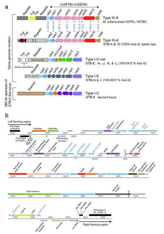
Global spread and limited genetic variation are hallmarks of M. tuberculosis, the agent of human tuberculosis. In contrast, Mycobacterium canettii and related tubercle bacilli that also cause human tuberculosis and exhibit unusual smooth colony morphology are restricted to East Africa. Here, we sequenced and analyzed the whole genomes of five representative strains of smooth tubercle bacilli (STB) using Sanger (4-5× coverage), 454/Roche (13-18× coverage) and/or Illumina DNA sequencing (45-105× coverage). We show that STB isolates are highly recombinogenic and evolutionarily early branching, with larger genome sizes, higher rates of genetic variation, fewer molecular scars and distinct CRISPR-Cas systems relative to M. tuberculosis. Despite the differences, all tuberculosis-causing mycobacteria share a highly conserved core genome. Mouse infection experiments showed that STB strains are less persistent and virulent than M. tuberculosis. We conclude that M. tuberculosis emerged from an ancestral STB-like pool of mycobacteria by gain of persistence and virulence mechanisms, and we provide insights into the molecular events involved.


Figures

Figure 1
Figure 1. Selection and genome features of analyzed strains
(a) multilocus sequence typing of 56 STB and 10 MTBC reference isolates. Phylogenetic positions based on split decomposition analysis of concatenated sequences of 12 house-keeping gene segments are represented. The scale bar represents Hamming distance. Numbers indicate the percent of bootstrap support of the splits obtained after 1,000 replicates. Arrows and stars indicate isolates selected for complete genome sequence and genome shotgun analyses, respectively. (b) pairwise, linear genomic comparisons of M. tuberculosis H37Rv, M. bovis AF2122/97, five selected STB strains, and two non-tuberculous mycobacterial species, M. marinum M and M. smegmatis mc2155. Red and blue lines indicate co-linear blocks of DNA:DNA similarity, and inverted matches, respectively. M. tub., M. tuberculosis; M. mar, M. marinum; M. smeg., M. smegmatis. (c) numbers of SNPs in pairwise comparisons between the indicated genomes. (d) Network phylogeny inferred among the five STB isolates subjected to complete genome sequence analysis and MTBC by NeighborNet analysis, based on pairwise alignments of whole genome SNP data. ‘*’ indicates 90% bootstrap support, while all other nodes had 100% support, 1000 iterations. (e) Histogram showing the respective numbers of SNPs between the aligned M. tuberculosis H37Rv and M. bovis or STB genomes (depicted in panel b).
Figure 2
Figure 2. CRISPR-Cas (clustered regularly interspaced short palindromic repeats-CRISPR-associated proteins) systems and prophages in STB and MTBC genomes
(a) Gene content of different CRISPR-Cas systems in MTBC and STB strains. Spacers are color-coded according to sequence similarities. Percentages of protein sequence identities are indicated between type III-A systems of M. tuberculosis H37Rv and STB A and D. The various combinations of identities between ubiquitous proteins (e.g. Cas2) of different CRISPR-Cas types are much lower (below 40%) and are not indicated. A star indicates a potential csb1 pseudogene in the system of STB-H. A broken line denotes ends of DNA sequence contigs variably delimiting the identified repeat zones of type I-E systems of STB-F, -G and -I. Mut id, mutual protein sequence identities; rep, repeats; Tnp, transposon; cas, csm, csb, csx, cse, various Cas gene families. (b) Schematic representation of a 55 kb spanning genomic region that encodes a putative prophage in STB strain I. STB-I genomic positions are marked on horizontal scales in bp. Brackets indicate a portion homologous to a prophage region in the M. marinum genome. Predicted coding sequences are shown above or below scales, corresponding to rightward and leftward transcription, respectively. Color-coding define features of predicted encoded products as follows. Gray, phage protein without database match or homologous to non-mycobacteriophage proteins of unknown function; blue, phage protein homologous to other mycobacteriophage proteins of unknown function (names of homologs are written in blue text, except for the portion homologous to the M. marinum prophage region); black, STB-I coding sequences and tRNA genes (conserved in other STB strains and M. tuberculosis H37Rv) flanking the phage insertion site corresponding to the Lys tRNA gene; all other colors, phage proteins with a predicted function (indicated in black text). A gray box on the second horizontal scale indicates a sequence contig break. Functional annotations of the predicted genes were made based on comparisons of the encoded products via the Genbank database, detection of protein domain signatures, and expert annotation of 374 other mycobacteriophage genomes retrieved from the PhagesDB database.
Figure 3
Figure 3. Inter-strain recombination segments between STB and MTBC genomes
(a) phylogenetic tree inferred by using Neighbor-Joining algorithm on nucleotide p-distances, after concatenation of sequence alignments of 2,047 genes of the predicted clonal portion of the STB-MTBC core genome (i.e. after exclusion of the genes affected by recombination- see text- and of gapped regions). (b) SNP distribution among STB and MTBC aligned genome segments, showing probable recombination regions involving genes rv1936-rv1937 between STB-J and M. tuberculosis. Each of the three panels shows a comparison of two STB or M. bovis strains (top, bottom) relative to M. tuberculosis H37Rv (middle). Red lines indicate individual SNPs identified between pairwise compared genomes. Thicker or uneven red lines result from multiple SNPs in close proximity or shifts due to small insertions/deletions. Note the SNP-free, identical genome segments between STB-J and H37Rv (boxed) conflict with their distant respective positions on the clonal core genome-based tree. M. tub. H37Rv, M. tuberculosis H37Rv.
Figure 4
Figure 4. Virulence and persistence of smooth tubercle bacilli (STB) and M. tuberculosis
(a) colony forming units (CFUs) recovered from lungs (a, c) and spleens (b, d) of BALB/c mice after intranasal infection with 103 CFUs. Panels a/b and c/d depict two independent experiments. The results are the median and range of CFUs from four mice. e, f, histopathological sections of lungs of BALB/c infected mice, 128 days post-intranasal infection with 103 CFUs. Blue circles show bronchi, “A” indicates alveoli, and “V” indicates blood-vessels.

References

    1. Dye C, Williams BG. The population dynamics and control of tuberculosis. Science. 2010;328:856–861. - PubMed
    1. Sreevatsan S, et al. Restricted structural gene polymorphism in the Mycobacterium tuberculosis complex indicates evolutionarily recent global dissemination. Proc Natl Acad Sci U S A. 1997;94:9869–9874. - PMC - PubMed
    1. Wirth T, et al. Origin, spread and demography of the Mycobacterium tuberculosis complex. PLoS Pathog. 2008;4:e1000160. - PMC - PubMed
    1. Brosch R, et al. A new evolutionary scenario for the Mycobacterium tuberculosis complex. Proc Natl Acad Sci U S A. 2002;99:3684–3689. - PMC - PubMed
    1. Supply P, et al. Linkage disequilibrium between minisatellite loci supports clonal evolution of Mycobacterium tuberculosis in a high tuberculosis incidence area. Mol Microbiol. 2003;47:529–538. - PubMed

Publication types

MeSH terms

LinkOut - more resources