Review
. 2013 May;22 Suppl 3(Suppl 3):S450-9.
doi: 10.1007/s00586-012-2626-z. Epub 2013 Jan 5.

Surgical management of primary spinal hemangiopericytomas: an institutional case series and review of the literature

Affiliations
Review

Surgical management of primary spinal hemangiopericytomas: an institutional case series and review of the literature

Ali Shirzadi et al. Eur Spine J. 2013 May.

Erratum in

  • Eur Spine J. 2013 May;22 Suppl 3:S460. Banykh, Sergei [corrected to Bannykh, Serguei];Patrick Johnson, J [corrected to Johnson, J P]

Abstract

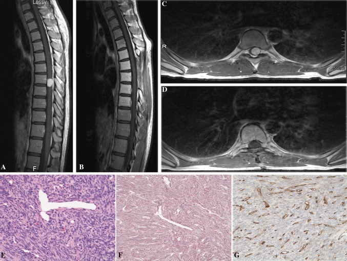
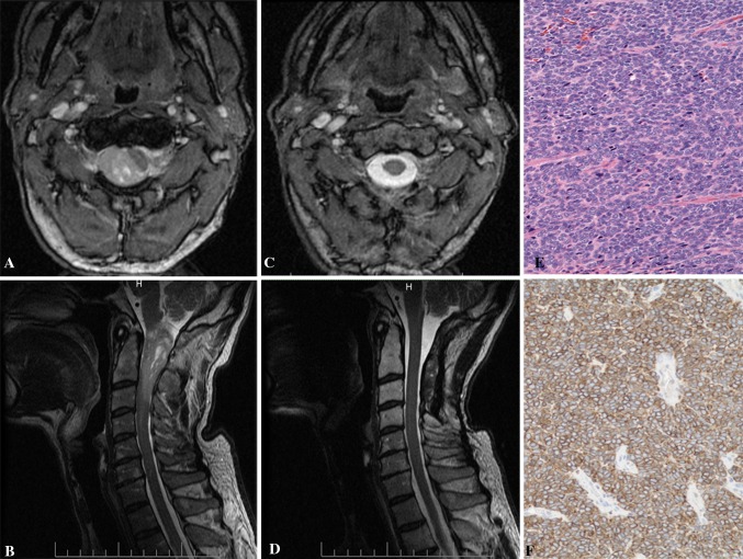
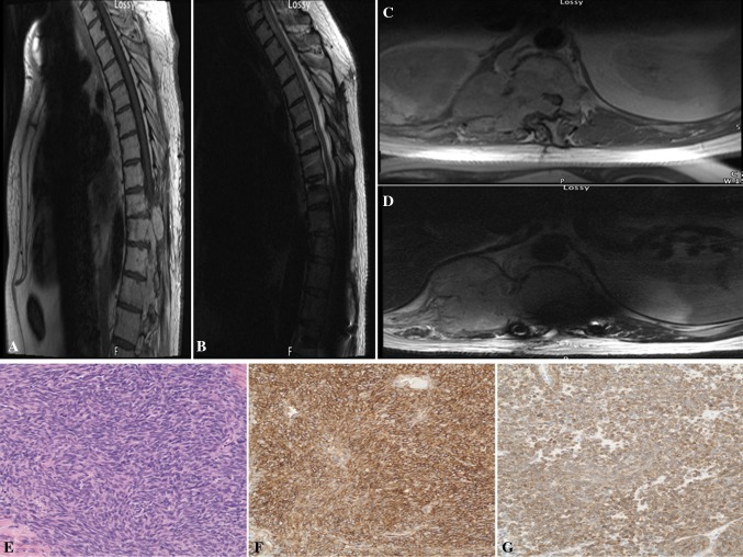
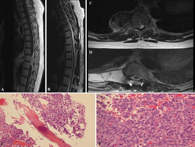
Purpose: Hemangiopericytoma (HPC) is a rare tumor of the central nervous system. Primary spinal occurrence of this tumor is extremely uncommon and cases involving the intramedullary spinal cord are even more rare. The purpose of this study was to explore the clinical features, surgical strategies, outcome and pathology in a consecutive series of patients treated at a single institution.

Methods: The authors performed a retrospective review of the clinicopathological characteristics of four patients with a pathological diagnosis of spinal HPC.

Results: Four cases with intradural as well as intra/extra-medullary components were identified. Gross total resection with no recurrence at the operative site was achieved in the majority of patients with a spinal HPC. One patient had significant recurrence and eventually, succumbed to the disease.

Conclusion: Increased awareness of these tumors' capability to occur intradurally and intramedullarly can help surgeons accurately diagnose and choose an effective plan of care. Gross total resection of hemangiopericytomas is the mainstay of treatment and should be pursued if feasible. Histopathology is essential to the diagnosis.


Figures

Fig. 1
Fig. 1
a, b Preoperative T1 sagittal and axial MRI with contrast showed a enhancing lesion at the T7–8 levels with severe compression of the spinal cord. c, d Postoperative T1 sagittal and axial MRI with contrast demonstrating resection of the tumor. e, f, g Hematoxylin and eosin (H&E) stained section showed moderately cellular proliferation of short nuclei, associated with prominent “staghorn” vasculature (e), embedded in a rich network of reticulin fibers (f), but showing only a weak staining for CD34 (g). Original magnification ×200
Fig. 2
Fig. 2
a, b Preoperative T1 sagittal and axial MRI with contrast showed a enhancing lesion at the C1–3 levels with severe compression of the spinal cord. c, d Postoperative T1 sagittal and axial MRI with contrast demonstrating resection of the tumor. e, f H&E stained sections disclosed tightly packed oval nuclei forming fascicles (e). Scattered tumor cells were positive for CD34 (f). Original magnification ×200
Fig. 3
Fig. 3
a, b Preoperative T1 sagittal and axial MRI with contrast showed a enhancing lesion at the T9–12 levels with severe compression of the spinal cord. c, d Postoperative T1 sagittal and axial MRI with contrast demonstrating recurrent tumor at T10–12 with complete obliteration of the spinal cord (d). e, f, g H&E stained sections revealed intersecting fascicles of tightly juxtaposed elongate tumor cells, associated with collagen bands (e). The tumor cells showed strong diffuse staining for CD34 and Bcl-2 (f, g, respectively). Original magnification ×200
Fig. 4
Fig. 4
a, b Preoperative T1 sagittal and axial MRI with contrast showed a enhancing lesion at the T9–10 levels with severe compression of the spinal cord. c, d Postoperative T1 sagittal and axial MRI with contrast demonstrating resection of the tumor. e, f There is focal bony involvement by tumor (e, H&E, 20×) and increased mitosis f, 40×)

References

    1. Stout AP, Murray MR. Hemanigopericytoma: a vascular tumor featuring Zimmermann’s pericytes. Ann Surg. 1942;116:26–33. doi: 10.1097/00000658-194207000-00004. - DOI - PMC - PubMed
    1. Giannini C, Rushing EJ, Hainfellner JA. Haemangiopericytoma. In: Louis DN, Ohgaki H, Wiestler OD, Cavenee WK, editors. WHO classification of tumours of the central nervous system. 4. Lyon: International Agency for Research on Cancer (IARC); 2007. pp. 178–180.
    1. Sundaram C, Shantveer UG, Megha US, Rekha SJ, Panigrahi KM, Purohit AK, Rammurti S. A clinicopathological and immunohistochemical study of the central nervous system hemangiopericytomas. J Clin Neurosci. 2010;17:469–472. doi: 10.1016/j.jocn.2009.08.018. - DOI - PubMed
    1. Ackerman PD, Khaldi A, Shea JF. Intradural hemangiopericytoma of the thoracic spine: a case report. Spine J. 2011;11(7):e9–e14. doi: 10.1016/j.spinee.2011.04.030. - DOI - PubMed
    1. Betchen S, Schwartz A, Black C, Post K. Intradural hemangiopericytoma of the lumbar spine: case report. Neurosurgery. 2002;50:654–657. - PubMed

LinkOut - more resources