. 2013 May;98(5):1009-27.
doi: 10.1113/expphysiol.2012.069369. Epub 2013 Jan 4.

The mechanical uncoupler blebbistatin is associated with significant electrophysiological effects in the isolated rabbit heart

Affiliations
Free PMC article

The mechanical uncoupler blebbistatin is associated with significant electrophysiological effects in the isolated rabbit heart

Kieran E Brack et al. Exp Physiol. 2013 May.
Free PMC article

Abstract

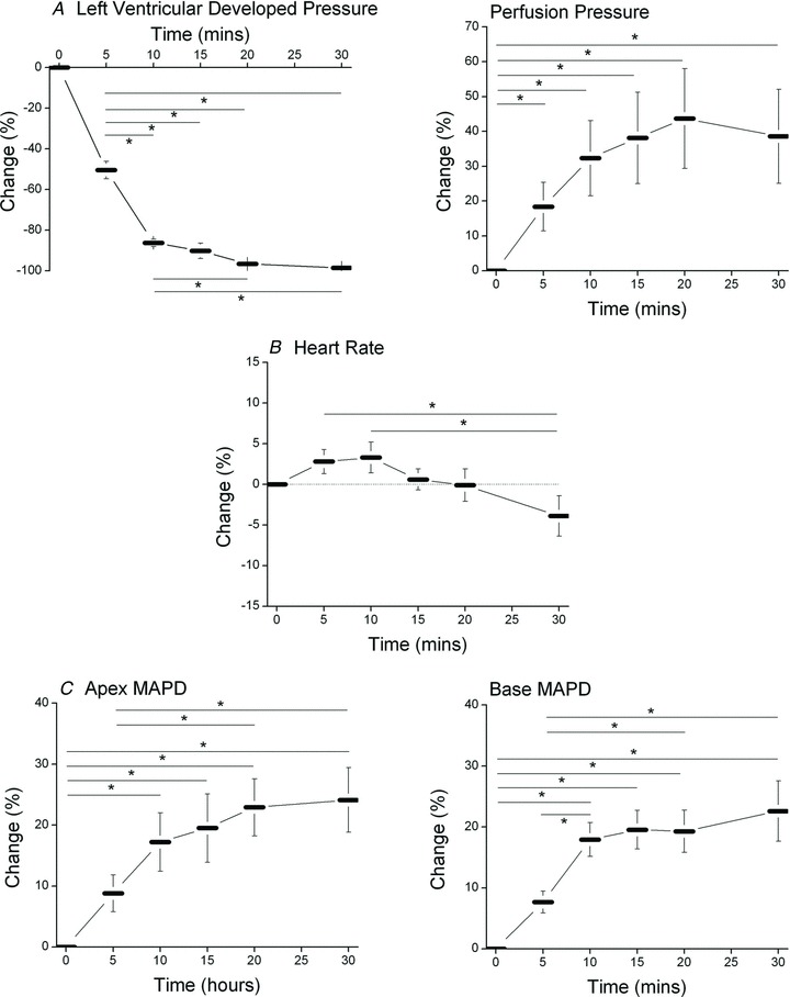
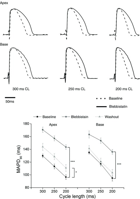
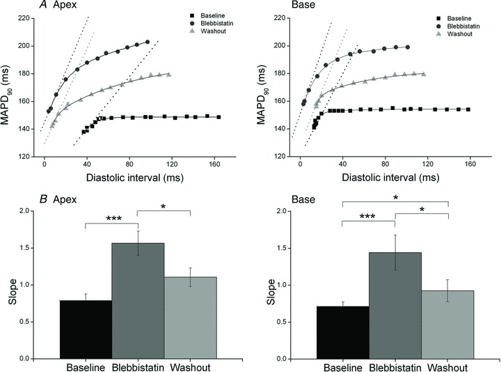
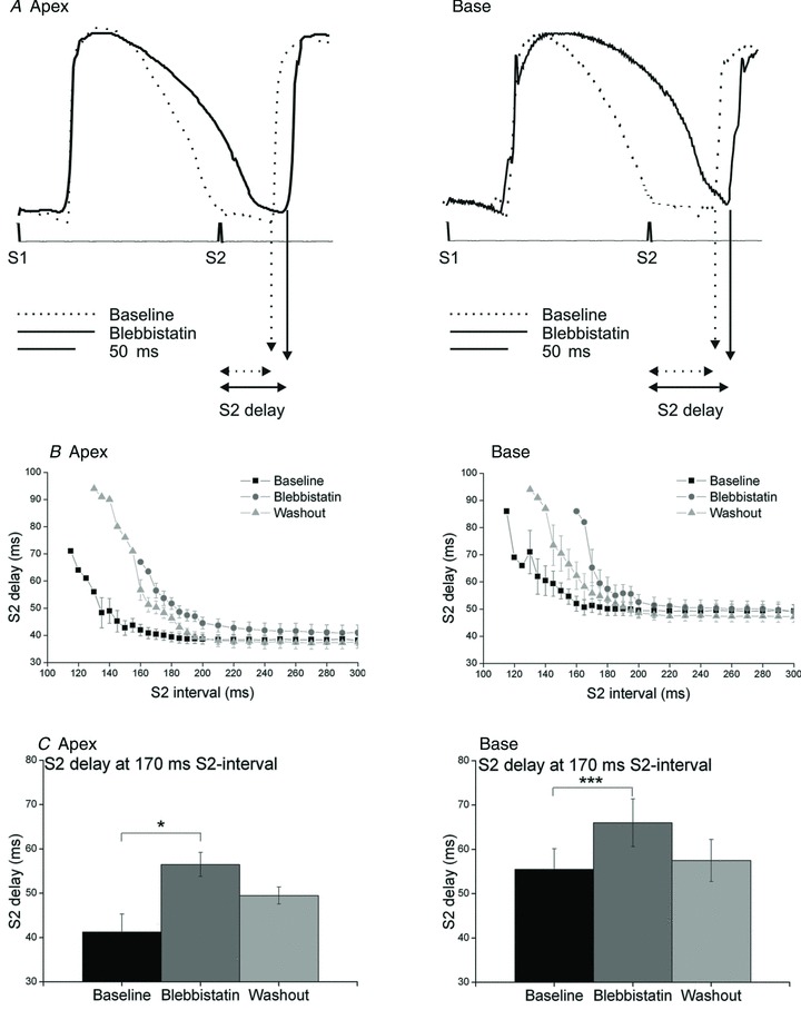
Blebbistatin (BS) is a recently discovered inhibitor of the myosin II isoform and has been adopted as the mechanical uncoupler of choice for optical mapping, because previous studies suggest that BS has no significant cardiac electrophysiological effects in a number of species. The aim of this study was to determine whether BS affects cardiac electrophysiology in isolated New Zealand White rabbit hearts. Langendorff-perfused hearts (n=39) in constant-flow mode had left ventricular monophasic action potential duration (MAPD) measured at apical and basal regions during constant pacing (300 ms cycle length). Standard action potential duration restitution was obtained using the single extrastimulus method with measurement of the maximal restitution slope. Ventricular fibrillation threshold was measured as the minimal current inducing sustained ventricular fibrillation with burst pacing (30 stimuli, at 30 ms intervals). Optical action potentials were recorded using the voltage-sensitive dye di-4-ANEPPS. Measurements were taken at baseline and after 60 min perfusion with BS (5 μm). Blebbistatin significantly prolonged left ventricular apical (mean±SEM; from 129.9±2.9 to 170.7±4.1 ms, P<0.001, n=8) and basal MAPD (from 135.0±2.3 to 163.3±5.6 ms, P<0.001) and effective refractory period (from 141.3±4.8 to 175.6±3.7 ms, P<0.001) whilst increasing the maximal slope of restitution (apex, from 0.79±0.09 to 1.57±0.16, P<0.001; and base, from 0.71±0.06 to 1.44±0.24, P<0.001) and ventricular fibrillation threshold (from 5.3±1.1 to 17.0±2.9 mA, P<0.001). In other hearts, blebbistatin significantly prolonged optically recorded action potentials (from 136.5±6.3 to 173.0±7.9 ms, P<0.05, n=4). In control experiments, the increase of MAPD with blebbistatin was present whether the hearts were perfused in constant-pressure mode (n=5) or in unloaded conditions (n=5). These data show that blebbistatin significantly affects cardiac electrophysiology. Its use in optical mapping studies should be treated with caution.


Figures

Figure 1
Figure 1. Time-based control data
Mean data illustrating repeated measurements of effective refractory period (ERP) and ventricular fibrillation threshold (A), monophasic action potential duration (MAPD) from the apex and base (B) and maximal slope of the electrical restitution curve at apical and basal sites (C) over a 5 h time period in the isolated, Langendorff-perfused rabbit heart (n= 8).
Figure 2
Figure 2. The effect of blebbistatin in the isolated rabbit heart
Raw data on the dynamic effect of blebbistatin (5 μm) on left ventricular pressure (LVP), perfusion pressure (PP), heart rate (HR) and monophasic action potential duration (MAPD) from the basal region in the isolated, Langendorff-perfused rabbit heart.
Figure 3
Figure 3. The dynamic effect of blebbistatin on cardiac physiology
Mean data showing the percentage change in left ventricular developed pressure and perfusion pressure (A), heart rate (B) and MAPD recorded from apical and basal sites (C) over the first 30 min perfusion with 5 μm blebbitstatin. *P < 0.05 as indicated by the horizontal bars, n= 8.
Figure 4
Figure 4. Effect of blebbistatin on MAPD
Top panel shows amplitude-normalized monophasic action potentials (MAPs) recorded from the apical (top traces) and basal region (bottom panel) of the left ventricle at baseline (dashed trances) and after 60 min of blebbistatin (continuous traces) during right ventricular pacing at cycle lengths (CL) of 300, 250 and 200 ms. Bottom panel, mean data showing MAPD90 at apical and basal sites at baseline, following 60 min perfusion with blebbistatin and following a 2 h washout period. *P < 0.05, ***P < 0.001, n= 8.
Figure 5
Figure 5. Effect of blebbistatin on action potential duration (APD) restitution
A, plot of APD restitution curves from a typical experiment at baseline, following 60 min of blebbistatin and following a 2 h washout period at apical (left) and basal regions (right), with the dotted lines representing the maximal slopes. Each symbol represents the APD at each S1–S2 interval, whilst the continuous line represents the fitted single exponential curve. B, mean data illustrating maximal restitution slope (Slope) at baseline, during perfusion with blebbistatin and after a 2 h period of washout at apal (left) and basal sites (right). *P < 0.05, ***P < 0.001, n= 8.
Figure 6
Figure 6. Effect of blebbistatin on S2 delay
A, raw data illustrating monophasic action potentials recorded at apical (left) and basal regions (right) at the S1–S2 coupling interval of 170 ms at baseline and during perfusion with blebbistatin. B, mean data (n= 8) illustrating S2 delay at baseline, after 60 min perfusion with blebbistatin and after a 2 h period of washout at apical (left) and basal regions (right). C, S2 delay at a selected S2 interval of 170 ms (n= 4).*P < 0.05, ***P < 0.001.
Figure 7
Figure 7. The effect of blebbistatin on the ECG
A, aligned and averaged ECGs. B, mean data recorded at baseline and at specific time points during perfusion with 5 μm blebbistatin. n= 7, *P < 0.05, **P < 0.01 versus time 0.
Figure 8
Figure 8. Control data
Mean data illustrating the effect of changing between constant-flow to constant-pressure Langendorff perfusion on MAPD (A), the effect of blebbistatin (Blebbi) perfusion on MAPD in hearts without any intraventricular load (unloaded; B) and the effect of blebbistatin in unloaded hearts during constant-pressure Langendorff perfusion (C). Experiments were conducted in our modified Tyrode solution (left panels) and in the Tyrode solution used by Fedorov et al. (2007; right panels; n= 5 in all groups). ***P < 0.001 versus baseline (BL)
Figure 9
Figure 9. The dose–response effect of blebbistatin
Left panels show aligned and averaged profiles for left ventricular pressure (LVP; A), perfusion pressure (PP; B), basal (C) and apical monophasic action potential duration (D) with 0.0, 0.5, 1, 5 and 10 μm blebbistatin perfusion. Right panels show mean data corresponding to each parameter. n= 6. *P < 0.05, **P < 0.01, ***P < 0.001 versus control. #P < 0.05, ##P < 0.01, ###P < 0.001 versus 0.5 μm blebbistatin. +P < 0.05 versus 1.0 μm blebbistatin.
Figure 10
Figure 10. Effect of blebbistatin on optical mapping recorded action potentials
Illustration of optically recorded action potentials using di-4-ANEPPS at base, mid-wall, free wall, apex and anterior wall sites at baseline and after perfusion with 5 μm blebbistatin during right ventricular pacing (300 ms cycle length).

References

    1. Baker LC, Wolk R, Choi BR, Watkins S, Plan P, Shah A, Salama G. Effects of mechanical uncouplers, diacetyl monoxime, and cytochalasin-D on the electrophysiology of perfused mouse hearts. Am J Physiol Heart Circ Physiol. 2004;287:H1771–H1779. - PubMed
    1. Biermann M, Rubart M, Moreno A, Wu J, Josiah-Durant A, Zipes DP. Differential effects of cytochalasin D and 2,3 butanedione monoxime on isometric twitch force and transmembrane action potential in isolated ventricular muscle: implications for optical measurements of cardiac repolarization. J Cardiovasc Electrophysiol. 1998;9:1348–1357. - PubMed
    1. Blanchard EM, Smith GL, Allen DG, Alpert NR. The effects of 2,3-butanedione monoxime on initial heat, tension, and aequorin light output of ferret papillary muscles. Pflugers Arch. 1990;416:219–221. - PubMed
    1. Brack KE, Coote JH, Ng GA. Modulation of ventricular fibrillation initiation and action potential duration restitution by acetylcholine, effect of varying extracellular calcium concentration. Cardiovasc Res. 2010;87(Suppl 1):S106–S106.
    1. Brack KE, Coote JH, Ng GA. Vagus nerve stimulation protects against ventricular fibrillation independent of muscarinic receptor blockade. Cardiovasc Res. 2011;91:437–446. - PubMed

Publication types

MeSH terms

Substances