. 2013 Apr;40(4):3083-92.
doi: 10.1007/s11033-012-2382-5. Epub 2013 Jan 5.

Assessment of atherosclerotic plaques in the rabbit abdominal aorta with interleukin-8 monoclonal antibody-targeted ultrasound microbubbles

Affiliations

Assessment of atherosclerotic plaques in the rabbit abdominal aorta with interleukin-8 monoclonal antibody-targeted ultrasound microbubbles

Yongping Lu et al. Mol Biol Rep. 2013 Apr.

Abstract

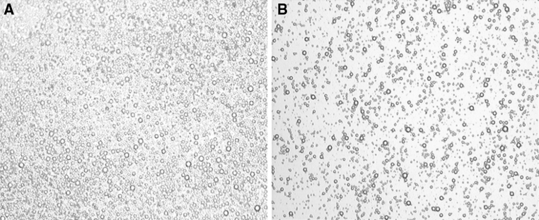
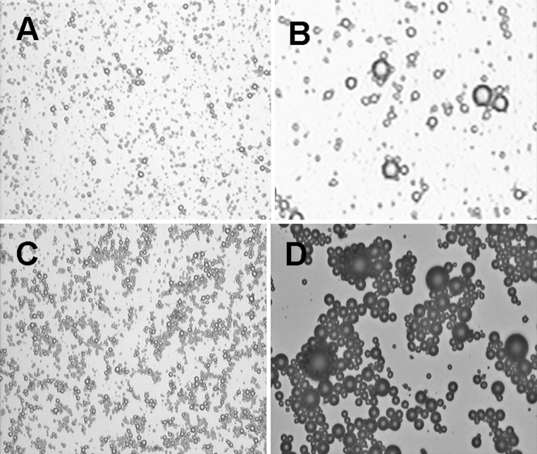
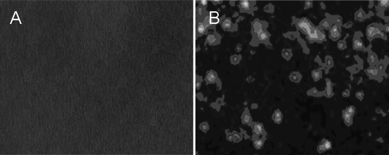
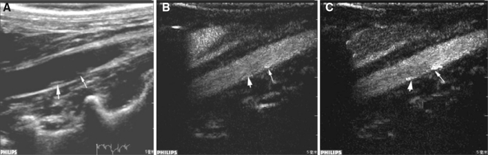
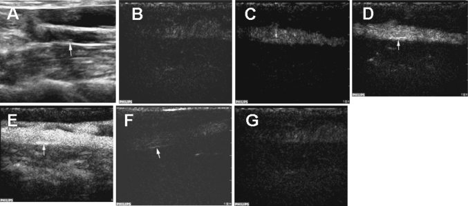
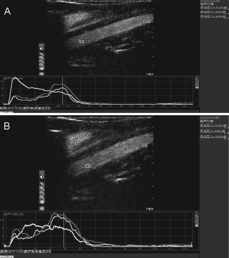
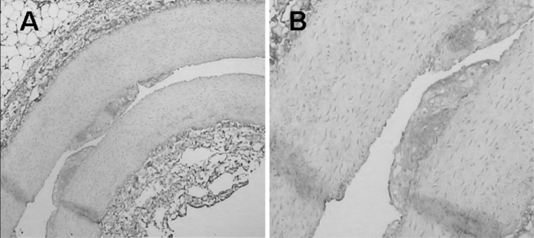
In this study, we aimed to prepare a neovascularization-relevant inflammatory cytokine-targeted ultrasound contrast agent and apply it in the ultrasound imaging of atherosclerotic plaque. An interleukin-8 (IL-8) monoclonal antibody was conjugated to SonoVue microbubbles using the N-succinimidyl-3-(2-pyridyldithio)propionate cross-linking method. Then, a prepared IL-8-targeted contrast agent was used for contrast-enhanced ultrasound (CEU) to detect rabbit abdominal aorta atherosclerotic plaque and to investigate the imaging characteristics of atherosclerotic plaque with the contrast agent. We found that an IL-8 monoclonal antibody can be successfully coupled to SonoVue microbubbles with stable biological characteristics. CEU with this IL-8-targeted contrast agent can increase the atherosclerotic plaque detection sensitivity, with stronger echo, so that three more plaques were detected compared with using non-targeted SonoVue microbubbles. Thus, an inflammatory cytokine-targeting ultrasound contrast agent carrying IL-8 monoclonal antibody can provide unique advantages for researching the characteristics of atherosclerotic plaque.


Figures

Fig. 1
Fig. 1
Light micrograph of SonoVue microbubbles. a Non-conjugated SonoVue microbubbles. b IL-8 monoclonal antibody-conjugated SonoVue microbubbles (×100)
Fig. 2
Fig. 2
Light micrograph of the mixture of SonoVue microbubbles and goat anti-mouse serum. a Non-conjugated microbubbles (×100). b Non-conjugated microbubbles (×400). c IL-8 monoclonal antibody-conjugated microbubbles (×100). d IL-8 monoclonal antibody-conjugated microbubbles (×400)
Fig. 3
Fig. 3
Immunofluorescence image of IgG-FITC-stained SonoVue microbubbles. a No fluorescence was observed on the surface of non-conjugated SonoVue microbubbles. b Bright-green fluorescence can be observed on the surface of IL-8-conjugated SonoVue microbubbles (×400). (Color figure online)
Fig. 4
Fig. 4
Representative ultrasound images of the atherosclerotic plaques in the rabbit abdominal aorta. a Two-dimensional image showing a plaque (large arrow); a soft plaque was invisible (small arrow). b Contrast ultrasound imaging with normal microbubbles showing two plaques with stronger echo compared with the two-dimensional view. The enhancement of the echo was more apparent for the soft plaque (small arrow). c Contrast ultrasound imaging with IL-8-targeted microbubbles showing two plaques with a markedly stronger echo compared with normal contrast ultrasound
Fig. 5
Fig. 5
Sequential ultrasound images of IL-8-targeted imaging for an atherosclerotic plaque of the rabbit abdominal aorta. a Conventional two-dimensional view. bg Sequential targeted CEU view: b 3.8 s post-contrast agent injection, c: 4.3 s post-contrast agent injection, d 4.9 s post-contrast agent injection; e 9.8 s post-contrast agent injection, f 68 s post-contrast agent injection, g 176 s post-contrast agent injection
Fig. 6
Fig. 6
The time-intensity curves of contrast ultrasound imaging with normal microbubbles (a) and IL-8-targeted microbubbles (b). The yellow, blue and red curves indicate the intensity of the lumen, intima and plaques, respectively. (Color figure online)
Fig. 7
Fig. 7
Typical histological images of rabbit abdominal aortic atherosclerotic plaque (a×100, b×400)
Fig. 8
Fig. 8
Immunohistochemical expression of IL-8 in atherosclerotic plaques in the rabbit abdominal aorta. Brown-stained area indicates positive IL-8 expression. (a ×40; b ×200). (Color figure online)
Fig. 9
Fig. 9
The missed soft plaque in two-dimensional images was identified by CD34 immunohistochemistry by its greatly increased brown staining (CD34-positive) in the inner part and base of the plaque compared with adjacent healthy intima. This result indicates a high neovascular density for the plaque, which is consistent with enrichment of the targeted microbubbles in contrast imaging. (a ×40; b ×100). (Color figure online)

References

    1. Abela GS, Picon PD, Friedl SE, Gebara OC, Miyamoto A, Federman M, Tofler GH, Muller JE. Triggering of plaque disruption and arterial thrombosis in an atherosclerotic rabbit model. Circulation. 1995;91(3):776–784. doi: 10.1161/01.CIR.91.3.776. - DOI - PubMed
    1. Apostolopoulos J, Davenport P, Tipping PG. Interleukin-8 production by macrophages from atheromatous plaques. Arterioscler Thromb Vasc Biol. 1996;16(8):1007–1012. doi: 10.1161/01.ATV.16.8.1007. - DOI - PubMed
    1. Cherkaoui S, Bettinger T, Hauwel M, Navetat S, Allémann E, Schneider M. Tracking of antibody reduction fragments by capillary gel electrophoresis during the coupling to microparticles surface. J Pharm Biomed Anal. 2010;53(2):172–178. doi: 10.1016/j.jpba.2010.01.039. - DOI - PubMed
    1. Dayton PA, Ferrara KW. Targeted imaging using ultrasound. J Magn Reson Imaging. 2002;16(4):362–377. doi: 10.1002/jmri.10173. - DOI - PubMed
    1. Ferrante EA, Pickard JE, Rychak J, Klibanov A, Ley K. Dual targeting improves microbubble contrast agent adhesion to VCAM-1 and P-selectin under flow. J Control Release. 2009;140(2):100–107. doi: 10.1016/j.jconrel.2009.08.001. - DOI - PMC - PubMed

Publication types

LinkOut - more resources