

Novel application of a spatial frequency domain imaging system to determine signature spectral differences between infected and noninfected burn wounds
- PMID: 23292572
- PMCID: PMC3539220
- DOI: 10.1097/BCR.0b013e318269be30
Novel application of a spatial frequency domain imaging system to determine signature spectral differences between infected and noninfected burn wounds
Abstract
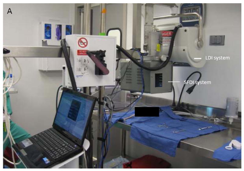
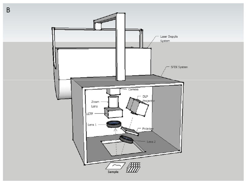
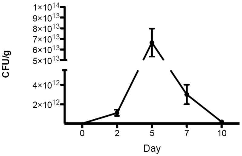
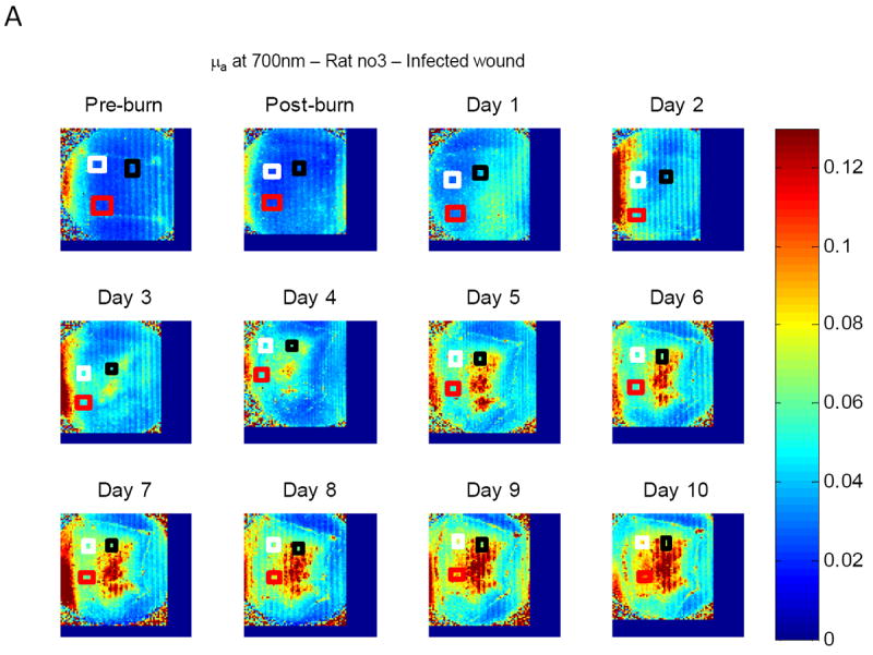
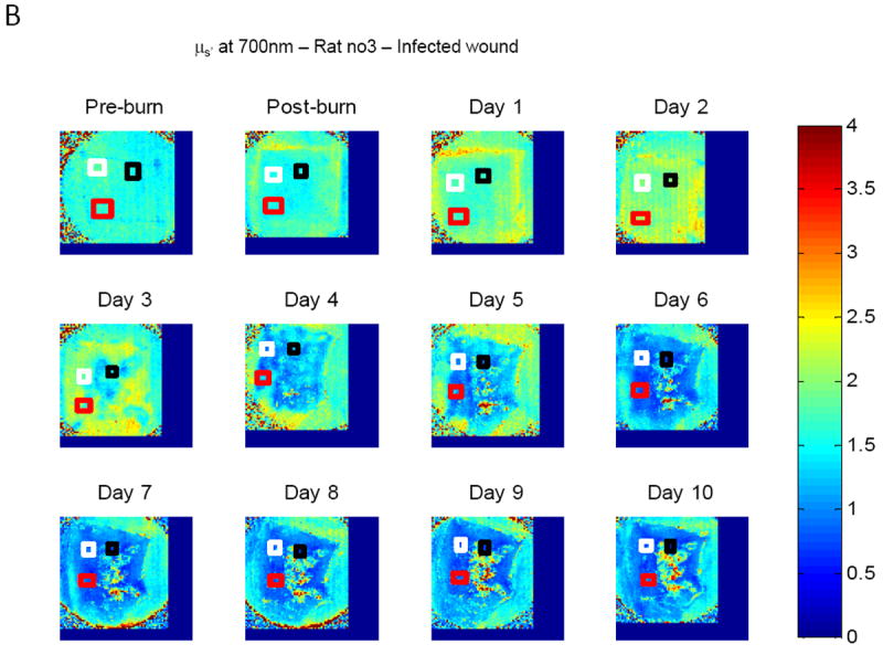
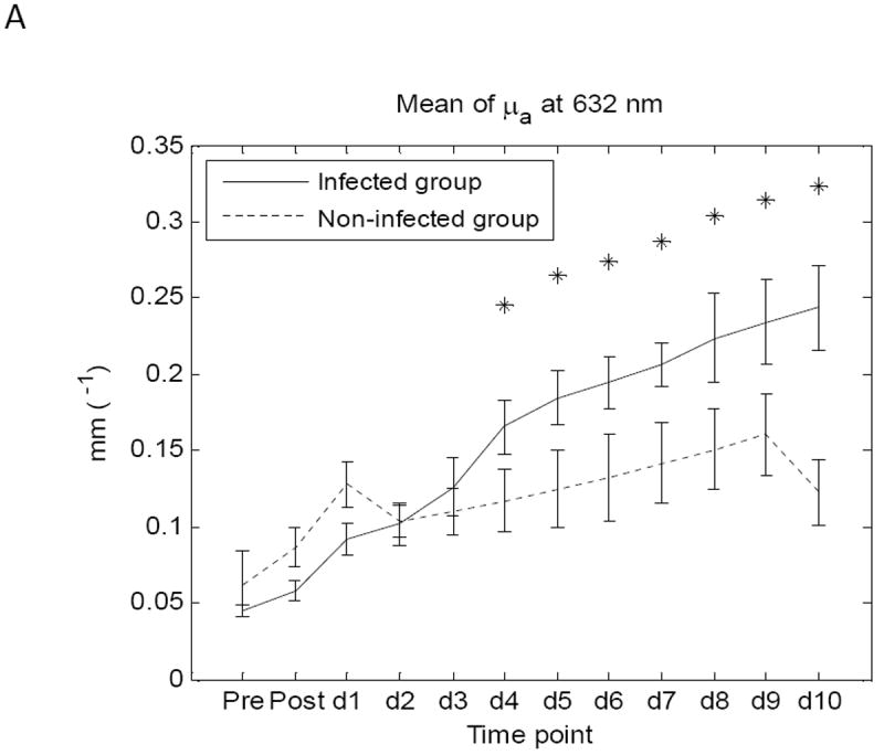
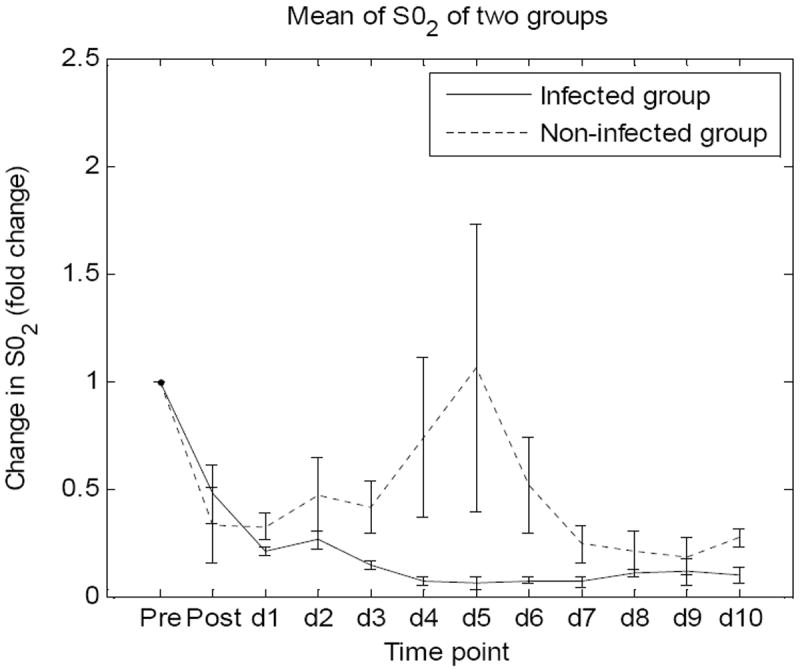
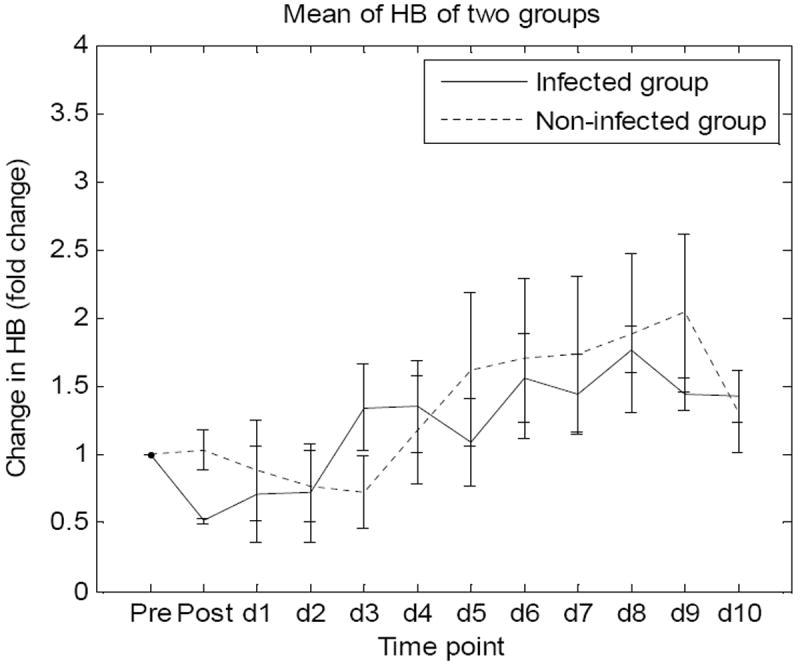
Complications of infection can increase burn-related morbidity and mortality. Early detection of burn wound infection could lead to more precise and effective treatment, reducing systemic complications and the need for long-term, broad-spectrum intravenous antibiotics. Quantitative cultures from biopsies are the accepted standard to determine infection. However, this methodology can take days to yield results and is invasive. This investigation focuses on the use of noninvasive imaging to determine the infection status of burn wounds in a controlled in vivo model. Full-thickness burn wounds were created on the dorsum of adult male rats (n = 6). Twenty-four hours after burn wound creation, wounds in the "Infected" group were inoculated with a vehicle containing 1 × 10(8) colony forming unit Staphylococcus aureus. "Control" group animals received vehicle alone. Subsequently, the wounds were imaged daily for a total of 10 days and the differences of skin optical properties were assessed using spatial frequency domain imaging at 16 different wavelengths from 500 to 700 nm. Regions of interest on the resulting images were selected and averaged at each time point. Statistically significant differences in average absorption and reduced scattering coefficients (μ(a) and μ(s)') at 620 and 700 nm were observed between the two groups (P < .05). Differential optical properties were most evident by day 4 and persisted throughout the time course. Differential signature changes in optical properties are evident in infected burn wounds. This novel application of spatial frequency domain imaging may prove to be a valuable adjunct to burn wound assessment. Further work will be aimed at determining dose-response relationships and prokaryotic species differences.
Figures

References
-
- Shurland S, et al. Comparison of mortality risk associated with bacteremia due to methicillin-resistant and methicillin-susceptible Staphylococcus aureus. Infect Control Hosp Epidemiol. 2007;28(3):273–9. - PubMed
-
- Kaiser ML, et al. Epidemiology and risk factors for hospital-acquired methicillin-resistant Staphylococcus aureus among burn patients. J Burn Care Res. 2011;32(3):429–34. - PubMed
-
- Schlievert PM, Kelly JA. Clindamycin-induced suppression of toxic-shock syndrome--associated exotoxin production. J Infect Dis. 1984;149(3):471. - PubMed
MeSH terms
Grants and funding
LinkOut - more resources
Full Text Sources
Other Literature Sources
Medical
Research Materials
