. 2013 Jan;25(1):215-28.
doi: 10.1105/tpc.112.106377. Epub 2013 Jan 4.

AUREOCHROME1a-mediated induction of the diatom-specific cyclin dsCYC2 controls the onset of cell division in diatoms (Phaeodactylum tricornutum)

Affiliations

AUREOCHROME1a-mediated induction of the diatom-specific cyclin dsCYC2 controls the onset of cell division in diatoms (Phaeodactylum tricornutum)

Marie J J Huysman et al. Plant Cell. 2013 Jan.

Abstract

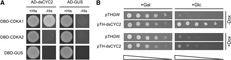
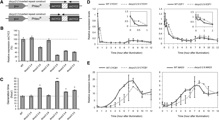
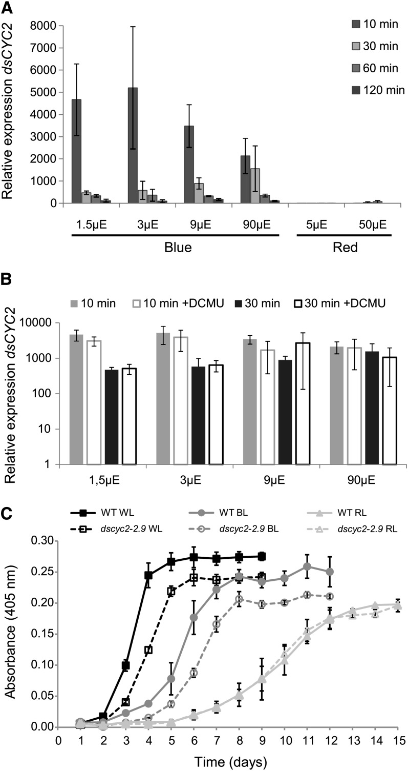
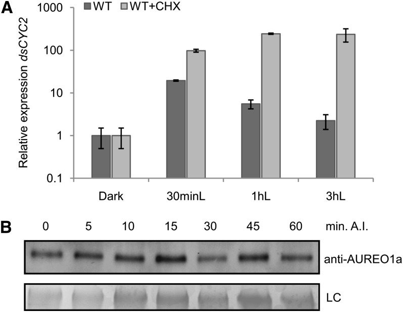
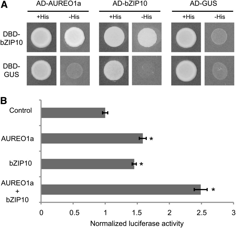
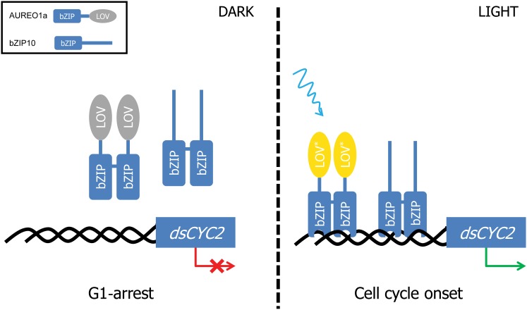
Cell division in photosynthetic organisms is tightly regulated by light. Although the light dependency of the onset of the cell cycle has been well characterized in various phototrophs, little is known about the cellular signaling cascades connecting light perception to cell cycle activation and progression. Here, we demonstrate that diatom-specific cyclin 2 (dsCYC2) in Phaeodactylum tricornutum displays a transcriptional peak within 15 min after light exposure, long before the onset of cell division. The product of dsCYC2 binds to the cyclin-dependent kinase CDKA1 and can complement G1 cyclin-deficient yeast. Consistent with the role of dsCYC2 in controlling a G1-to-S light-dependent cell cycle checkpoint, dsCYC2 silencing decreases the rate of cell division in diatoms exposed to light-dark cycles but not to constant light. Transcriptional induction of dsCYC2 is triggered by blue light in a fluence rate-dependent manner. Consistent with this, dsCYC2 is a transcriptional target of the blue light sensor AUREOCHROME1a, which functions synergistically with the basic leucine zipper (bZIP) transcription factor bZIP10 to induce dsCYC2 transcription. The functional characterization of a cyclin whose transcription is controlled by light and whose activity connects light signaling to cell cycle progression contributes significantly to our understanding of the molecular mechanisms underlying light-dependent cell cycle onset in diatoms.


Figures

Figure 1.
Figure 1.
Light-Dependent Transcription and Translation of dsCYC2. (A) Schematic representation of the HA marker construct. (B) Transcript levels of dsCYC2 (solid line) and HA-tagged dsCYC2 (dashed line) during a 60-min time course after illumination (A.I.) of 24-h dark-adapted HA marker cells. Values were normalized against those obtained for histone H4 and then rescaled to the gene expression levels at 0 min after illumination (=1). Error bars represent se of three technical replicates. (C) dsCYC2-HA protein levels during a 12-h (top panel) and 60-min (bottom panel) time course after illumination of 24-h dark-adapted HA marker cells. −, Negative control (wild-type 4 h light); +, positive control (HA 4 h light). LC, loading control by Coomassie blue staining.
Figure 2.
Figure 2.
dsCYC2 Functions as a G1-Cyclin. (A) Interaction of dsCYC2 with CDKA1. Yeast PJ694-α cells were cotransformed with bait and prey plasmid as indicated. Cotransformation was analyzed on medium lacking Leu and Trp (+His). Cotransformants were tested for their ability to activate the His marker gene by assessing yeast growth on medium lacking Leu, Trp, and His (-His). Constructs containing β-glucuronidase (GUS) were used as negative controls. For each combination, three independent colonies were screened, one of which is shown. (B) Complementation of G1 cyclin–deficient yeast by dsCYC2. BF305-15d-21 cells were transformed with pTHGW (vector control) or pTH-dsCYC2. Yeast cells were serially diluted and spotted onto SD-Ura plates containing Gal (+Gal) or Glc (+Glc). When Glc was the sole carbon source, control cells were not able to grow because of the lack of G1 cyclin expression, while cells that expressed dsCYC2 overcame this phenotype. When dsCYC2 expression was repressed by the addition of doxycycline (+Dox), the complementation was lost.
Figure 3.
Figure 3.
Effect of dsCYC2 Silencing on Cell Cycle Progression. (A) Schematic representation of the dsCYC2 inverted repeat constructs used for silencing analysis. In the dscyc2-2 construct, the large fragment is positioned first, followed by the small fragment. In the dscyc2-3 construct, the small fragment is followed by the large fragment (arrows). (B) Real-time quantitative PCR analysis of dsCYC2 transcript levels in wild-type (WT) and silenced lines. Cells were dark adapted for 24 h, and transcript levels were measured 15 min after light exposure. Transcript levels of wild-type cells were set at 100%. (C) Generation times of wild-type and dsCYC2 silenced lines grown at 100 µE 12L/12D cycles. Error bars (in (B) and (C) represent sd of the mean of three independent experiments. *P < 0.005; **P < 0.001 (two-tailed Student’s t test). (D) and (E) Transcript expression profiles of G1 marker genes (D) and mitotic markers (E) during a synchronized time course in wild-type and dscyc2-2.9 knockdown cells. Error bars represent se of two biological replicates.
Figure 4.
Figure 4.
Blue Light Photoreceptor-Mediated Control of dsCYC2 Induction. (A) Wavelength and fluence rate dependency of dsCYC2 induction. Wild-type cultures were dark incubated for 60 h and switched to blue or red light at different light intensities, as indicated. Relative mRNA levels of dsCYC2 at 10, 30, 60, and 120 min after light exposure are shown. Relative levels were normalized to histone H4 levels and rescaled to the expression level in dark-incubated cells (=1). (B) Effect of DCMU on dsCYC2 induction. Log scale representation of the relative mRNA levels of dsCYC2 at 10 and 30 min after blue light exposure at different light intensities in the absence or presence of DCMU. In (A) and (B), error bars represent se of two biological replicates. (C) Growth curves of wild-type (WT) and dscyc2-2.9 cells grown in white (WL), blue (BL), and red (RL) light adjusted to equal values of photosynthetically absorbed radiation. Error bars represent sd of three biological replicates.
Figure 5.
Figure 5.
Regulation of the dsCYC2 Promoter by Light. (A) Schematic representation of the prom-eYFP marker (right) constructs. (B) Transcript levels of dsCYC2 and eYFP during a 60-min time course after illumination (A.I.) of 24-h dark-adapted pdsCYC2-eYFP cells. Values were normalized against H4 expression levels and rescaled to the levels at 0 min after illumination (= 1). Error bars represent se of two biological replicates. (C) Y1H protein-DNA interaction assay. Interactions are positive when HIS3 (growth on 3-aminotriazole–containing medium (+3AT) and LacZ (X-Gal turns blue) expression is induced. Constructs containing GUS were used as negative controls. For each combination, three independent colonies were screened, one of which is shown.
Figure 6.
Figure 6.
Posttranslational Regulation of dsCYC2 Induction upon Light Exposure. (A) Wild-type (WT) cultures were synchronized by 24-h dark treatment (Dark) and then exposed to light for 0.5 (30 minL), 1 (1 hL), or 3 h (3 hL) in the absence (dark gray) or presence (light gray) of 2 μg/mL CHX. Relative expression levels of dsCYC2 are shown. Values were normalized against H4 expression levels and then rescaled to the gene expression levels of the dark sample (=1). Error bars represent se of two independent experiments. (B) AUREO1a protein levels during a 60-min time course after illumination (A.I.) of 24-h dark-adapted HA marker cells. LC, loading control by Coomassie blue staining.
Figure 7.
Figure 7.
Activation of the dsCYC2 Promoter by AUREO1a and bZIP10. (A) Y2H protein–protein interaction assay. Yeast cells were cotransformed with bait and prey plasmid as indicated. Cotransformation was analyzed on medium lacking Leu and Trp (+His). Cotransformants were tested for their ability to activate the His marker gene by assessing yeast growth on medium lacking Leu, Trp, and His (-His). Constructs containing GUS were used as negative controls. For each combination, three independent colonies were screened, one of which is shown. (B) Protoplast transactivation assay using pdsCYC2:fLUC as reporter, p35S:rLUC as normalization, and p35S:AUREO1a and p35S:bZIP10 as effector constructs. Luciferase activity of the control was arbitrarily set to 1. Error bars represent se of three biological replicates (*P ≤ 0.05, two-sided t test).
Figure 8.
Figure 8.
Hypothetical Model of the Light-Dependent Regulation of dsCYC2 and Cell Cycle Onset in P. tricornutum. Upon light exposure, the LOV domain of AUREO1a is changed from the dark state (gray) into the light state (yellow) through cysteinyl-FMN adduct formation. This induces a conformational change in the homodimer protein complex, resulting in the binding of the bZIP domains to the promoter of dsCYC2. Binding of both AUREO1a and bZIP10 homodimers to different regulatory elements in the dsCYC2 promoter results in the synergistic activation of dsCYC2 and leads to the onset of the cell cycle.

References

    1. Bailleul B., Rogato A., de Martino A., Coesel S., Cardol P., Bowler C., Falciatore A., Finazzi G. (2010). An atypical member of the light-harvesting complex stress-related protein family modulates diatom responses to light. Proc. Natl. Acad. Sci. USA 107: 18214–18219 - PMC - PubMed
    1. Bisova K., Krylov D.M., Umen J.G. (2005). Genome-wide annotation and expression profiling of cell cycle regulatory genes in Chlamydomonas reinhardtii. Plant Physiol. 137: 475–491 - PMC - PubMed
    1. Bowler C., et al. (2008). The Phaeodactylum genome reveals the evolutionary history of diatom genomes. Nature 456: 239–244 - PubMed
    1. Bradford M.M. (1976). A rapid and sensitive method for the quantitation of microgram quantities of protein utilizing the principle of protein-dye binding. Anal. Biochem. 72: 248–254 - PubMed
    1. Brzezinski M.A., Olson R.J., Chisholm S.W. (1990). Silicon availability and cell-cycle progression in marine diatoms. Mar. Ecol. Prog. Ser. 67: 83–96

Publication types

LinkOut - more resources