. 2013 Mar;195(5):1090-9.
doi: 10.1128/JB.01344-12. Epub 2013 Jan 4.

Novel twin streptolysin S-like peptides encoded in the sag operon homologue of beta-hemolytic Streptococcus anginosus

Affiliations

Novel twin streptolysin S-like peptides encoded in the sag operon homologue of beta-hemolytic Streptococcus anginosus

Atsushi Tabata et al. J Bacteriol. 2013 Mar.

Abstract

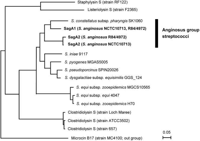
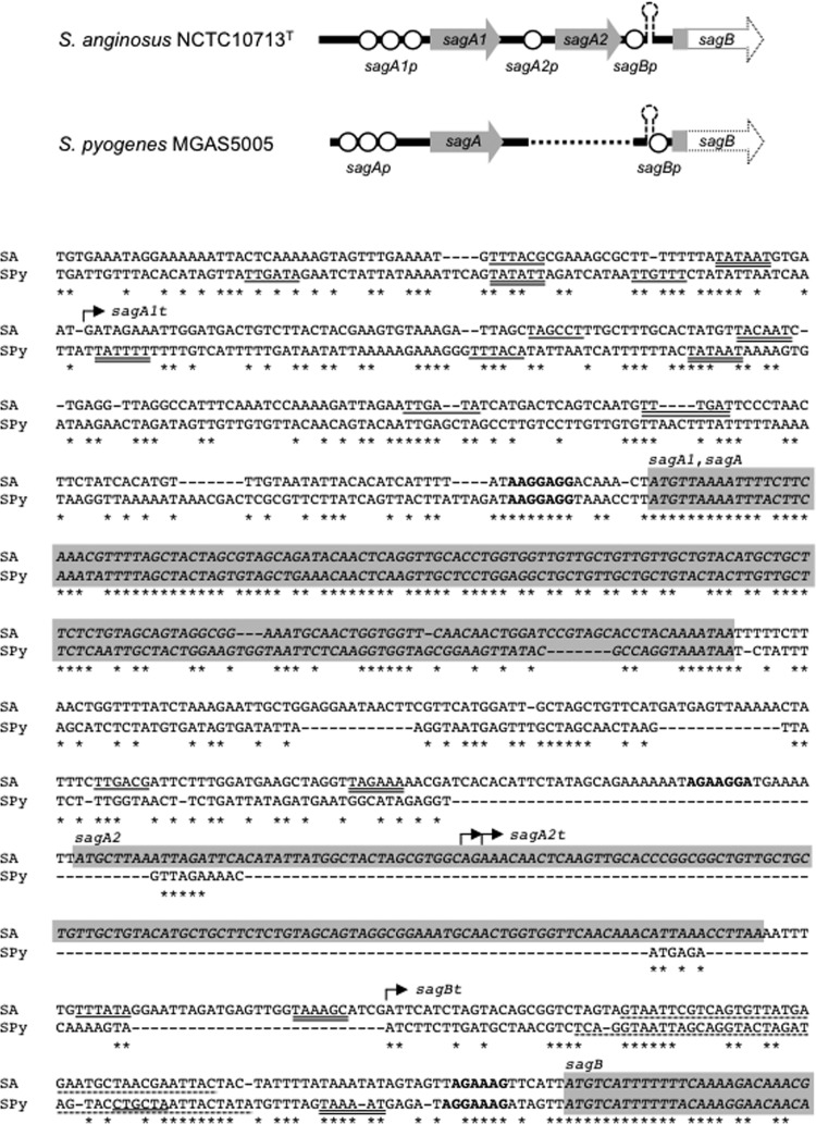
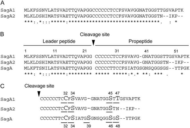
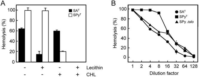
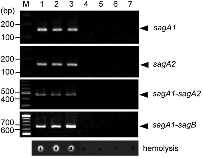
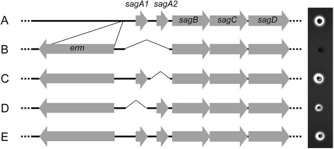
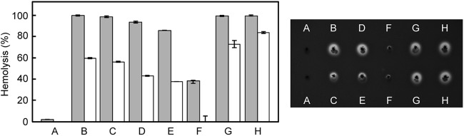
Streptococcus anginosus is a member of the anginosus group streptococci, which form part of the normal human oral flora. In contrast to the pyogenic group streptococci, our knowledge of the virulence factors of the anginosus group streptococci, including S. anginosus, is not sufficient to allow a clear understanding of the basis of their pathogenicity. Generally, hemolysins are thought to be important virulence factors in streptococcal infections. In the present study, a sag operon homologue was shown to be responsible for beta-hemolysis in S. anginosus strains by random gene knockout. Interestingly, contrary to pyogenic group streptococci, beta-hemolytic S. anginosus was shown to have two tandem sagA homologues, encoding streptolysin S (SLS)-like peptides, in the sag operon homologue. Gene deletion and complementation experiments revealed that both genes were functional, and these SLS-like peptides were essential for beta-hemolysis in beta-hemolytic S. anginosus. Furthermore, the amino acid sequence of these SLS-like peptides differed from that of the typical SLS of S. pyogenes, especially in their propeptide domain, and an amino acid residue indicated to be important for the cytolytic activity of SLS in S. pyogenes was deleted in both S. anginosus homologues. These data suggest that SLS-like peptides encoded by two sagA homologues in beta-hemolytic S. anginosus may be potential virulence factors with a different structure essential for hemolytic activity and/or the maturation process compared to the typical SLS present in pyogenic group streptococci.


Figures

Fig 1
Fig 1
Genetic structure of sagSA (A) and the “typical” sag operon from other pyogenic group streptococci (B). sagSA is characterized by the presence of twin sagA homologues (sagA1 and sagA2). The arrows in panel A indicate the insertion sites of pGhost9:ISS1 in four nonhemolytic transformants obtained in random gene knockout experiments.
Fig 2
Fig 2
Phylogenetic tree of SagA of streptococci and their homologues in other bacterial genera. The amino acid sequences were aligned by ClustalX, and the phylogenetic tree was constructed by Njplot using microcin B17 as an out-group. SagA1 and SagA2 from S. anginosus strains are indicated in boldface.
Fig 3
Fig 3
Genetic structure of the promoter regions of sagA1 to sagB in S. anginosus and sagA to sagB in S. pyogenes. The typical prokaryotic promoters predicted from their DNA sequences are shown with suffix “p” in the scheme at the top. The nucleotide sequence alignments of the region from the promoter of sagA1 to the head part of sagB ORF of S. anginosus NCTC10713T (SA) and from the promoter of sagA to the head part of sagB of S. pyogenes MGAS5005 (SPy) are also shown. The deduced −35 and −10 regions are single and double underlined, respectively. The potential promoters with a score value above 57.00 predicted in the analyses using the program “Search for Promoter Sequence” within GENETYX software are shown. Moreover, a deduced promoter for sagB of S. anginosus is also shown, although the score value in this case was 53.25. Rho-independent terminators are indicated by a broken line, and SD sequences shown in bold. The ORF of each gene is shown italicized with a shaded background. The start point of each transcript of sagSA revealed by 5′-RACE analysis (sagA1t, sagA2t, and sagBt) is indicated by an arrow.
Fig 4
Fig 4
Alignments of amino acid sequences of SagA and homologues. (A) Identity of SagA1 with SagA2 of S. anginosus NCTC10713T. (B) Alignment of the amino acid sequences of SagA homologues: SagA1 and SagA2 of S. anginosus NCTC10713T and typical SagA of S. pyogenes MGAS5005. The alignment analyses were conducted using ClustalX. The symbols indicating identity/homology of primary sequence are as follows: *, identical amino acid, “:,” amino acid highly conserved; “.,” amino acid somewhat similar. The amino acids deduced to contribute to heterocycle formation in SagA1, SagA2, and SagA are underlined, and the amino acids predicted to be indispensable residues for heterocycle formation according to the analysis in SagA are shown in large font (C).
Fig 5
Fig 5
(A) 5′-RACE analysis of sagSA. Total RNA from early-logarithmic-phase cells was prepared and used for 5′-RACE analysis. (A) The resulting DNA fragments containing the 5′ terminus of cDNA from mRNA sequence(s) of sagSA were amplified by nested PCR and analyzed by agarose-gel electrophoresis. M, hundred-base-pair ladder marker. (B) Cloned 5′-RACE products for sequencing. The 5′-RACE product cloned in the SmaI site of pUC18 was obtained by double digestion with EcoRI and SalI and separated by agarose gel electrophoresis. M, hundred-base-pair ladder size marker. Lanes 1 to 4 show the 5′-end part of cDNA deduced to be the transcripts from sagA1p (lane 1), sagA2p (lanes 2 and 3), and sagBp (lane 4).
Fig 6
Fig 6
(A) Effect of lecithin and cholesterol on hemolysis induced by the hemolysin secreted by S. anginosus and S. pyogenes type strains. Culture supernatants were preincubated with 0.02% (wt/vol) lecithin or 1 μM cholesterol (CHL) and then incubated with human erythrocytes. Control reaction mixtures contained no lecithin or cholesterol. All results are shown as mean values (n = 3) with standard deviations (SD). (B) Dose dependency of hemolysis by culture supernatant of the S. anginosus type strain, S. pyogenes type strain, and the S. pyogenes slo-deleted mutant (Δslo). The supernatants from mid- to late-logarithmic-phase culture (OD660 of 1.0) of the S. anginosus type strain (circles), S. pyogenes type strain (squares), and the S. pyogenes Δslo mutant (triangle) were 2-fold serially diluted and then incubated with human erythrocytes. All results are shown as mean values (n = 2) with differences.
Fig 7
Fig 7
Relationship between the presence of the sagA1 and sagA2 genes and beta-hemolysis. Typical results of PCR amplification obtained in three beta-hemolytic strains and four nonhemolytic strains of S. anginosus are shown. Beta-hemolysis on blood agar plates for these strains is also indicated.
Fig 8
Fig 8
Relationship between the presence of sagA homologues and beta-hemolysis from gene deletion experiments. Schemes of the genetic structure of parent strain S. anginosus NCTC10713T (A), the sagA1 sagA2 double-deletion mutant (B), the sagA2 single-deletion mutant (C), the sagA1 single-deletion mutant (D), and a control mutant containing an erythromycin resistance gene cassette in the reverse direction just upstream of the intact sagSA (E) are shown together with their beta-hemolytic activity on human blood agar plates.
Fig 9
Fig 9
Complementation of sagA homologues in the sagA1 sagA2 double-deletion mutant. (Right) Hemolysis on human blood agar after 1 day of cultivation. (A) The sagA1 sagA2 double-deletion mutant. (B to F) Complementation of strains with both sagA1 and sagA2 transcribed by the intact promoter of sagA1 with three potential promoters (Fig. 3) (B), by the truncated promoter of sagA1 with the second and the last potential promoters (see Fig. 3) (C), with only sagA1 transcribed by the intact promoter of sagA1 (D) or by the truncated promoter of sagA1 (E), or with only sagA2 transcribed by its own promoter (F). Hemolytic patterns of a control mutant with an erythromycin-resistance gene cassette just upstream of intact sagSA (G) and the parent strain (H) are also shown. (Left) Graph showing the hemolytic activity of the culture supernatant for each strain. Gray bars indicate the hemolytic activity of the original culture supernatant, and white bars indicate the hemolytic activity of culture supernatant diluted 3-fold with PBS. Results are shown as mean values (n = 2) with the differences between duplicates indicated.

References

    1. Whiley RA, Beighton D. 1991. Emended descriptions and recognition of Streptococcus constellatus, Streptococcus intermedius, and Streptococcus anginosus as distinct species. Int. J. Syst. Bacteriol. 41:1–5 - PubMed
    1. Whiley RA, Hall LMC, Hardie JM, Beighton D. 1999. A study of small-colony, β-hemolytic, Lancefield group C streptococci within the anginosus group: description of Streptococcus constellatus subsp. pharynges subsp. nov., associated with the human throat and pharyngitis. Int. J. Syst. Bacteriol. 49:1443–1449 - PubMed
    1. Whiley RA, Beighton D, Winstanley TG, Fraser HY, Hardie JM. 1992. Streptococcus intermedius, Streptococcus constellatus, and Streptococcus anginosus (the Streptococcus milleri group): association with different body sites and clinical infections. J. Clin. Microbiol. 30:243–244 - PMC - PubMed
    1. Ruoff KL. 1988. Streptococcus anginosus (“Streptococcus milleri”): the unrecognized pathogen. Clin. Microbiol. Rev. 1:102–108 - PMC - PubMed
    1. Clarridge JE, III, Attorri S, Musher DM, Hebert J, Dunber S. 2001. Streptococcus intermedius, Streptococcus constellatus, and Streptococcus anginosus (“Streptococcus milleri group”) are of different clinical importance and are not equally associated with abscess. Clin. Infect. Dis. 32:1511–1515 - PubMed

MeSH terms

Associated data

LinkOut - more resources