

Molecular testing prognostic of low risk in epithelioid uveal melanoma in a child
- PMID: 23292925
- PMCID: PMC4966167
- DOI: 10.1136/bjophthalmol-2012-302561
Molecular testing prognostic of low risk in epithelioid uveal melanoma in a child
Abstract
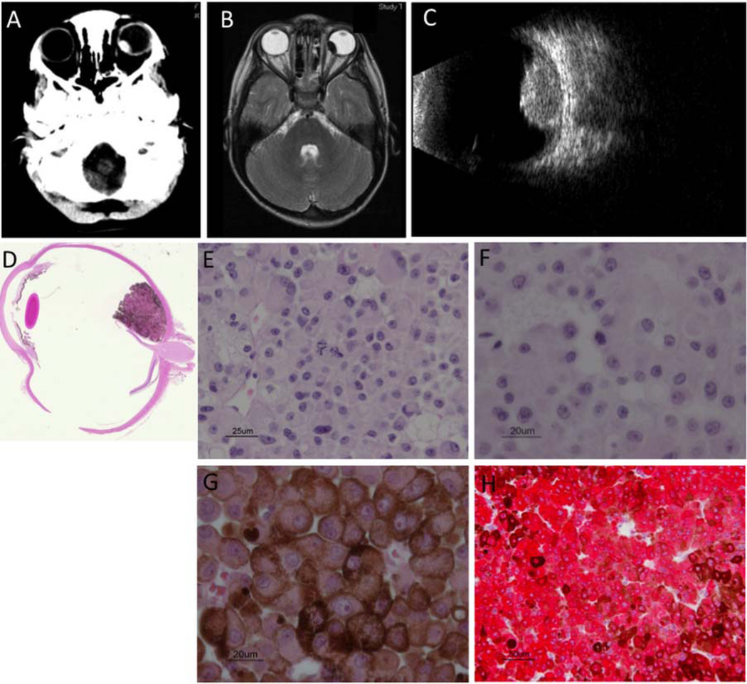
Aims: To characterise a histologically unusual paediatric uveal melanoma by gene expression and karyotypic profiling and assess prognosis.
Methods: The tumour was studied by histopathology, karyotype analysis, single nucleotide polymorphism and gene expression profile analysis for correlation with clinical outcome.
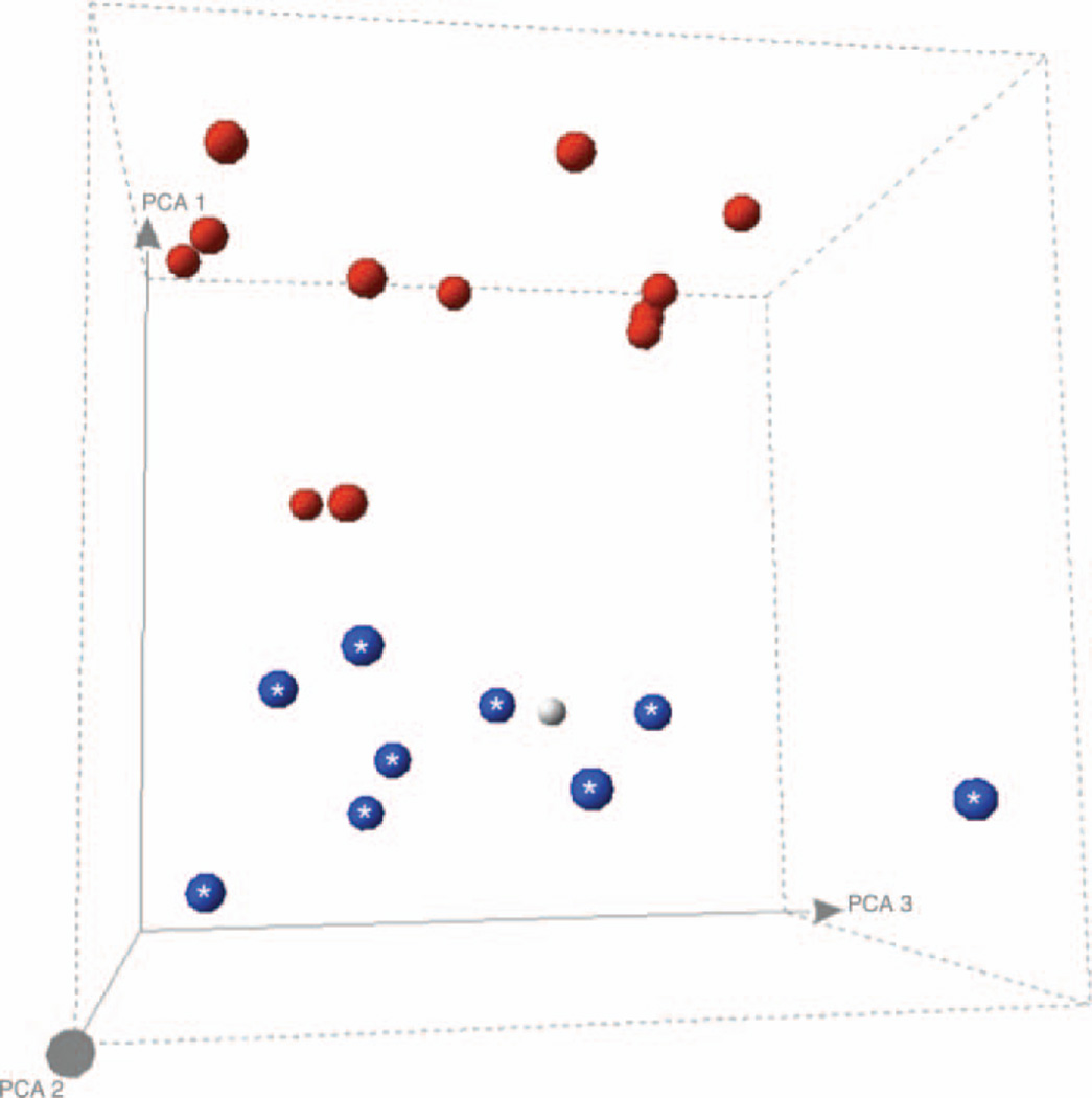
Results: The tumour had predominantly epithelioid histology. Karyotype analysis showed none of the poor prognosis features normally associated with uveal melanoma. single nucleotide polymorphism analysis revealed no imbalance at chromosome 3. Gene expression profiling indicated low risk disease.
Conclusions: We report a child remaining relapse-free 6 years after diagnosis of a very rare uveal melanoma, with poor prognosis epithelioid histology, but gene expression profiling that accurately predicted low risk disease.
Figures
References
-
- Singh AD, Shields CL, Shields JA, et al. Uveal melanoma in young patients. Arch Ophthalmol. 2000;118:918–923. - PubMed
-
- Kanthan GL, Grigg J, Billson F, et al. Paediatric uveal melanoma. Clin Experiment Ophthalmol. 2008;36:374–376. - PubMed
-
- Shields CL, Kaliki S, Shah SU, et al. Iris melanoma: features and prognosis in 317 children and adults. J Aapos. 2012;16:10–16. - PubMed
-
- Barr CC, McLean IW, Zimmerman LE. Uveal melanoma in children and adolescents. Arch Ophthalmol. 1981;99:2133–2136. - PubMed
-
- Kilic E, van Gils W, Lodder E, et al. Clinical and cytogenetic analyses in uveal melanoma. Invest Ophthalmol Vis Sci. 2006;47:3703–3707. - PubMed
Publication types
MeSH terms
Grants and funding
LinkOut - more resources
Full Text Sources
Other Literature Sources
Medical