Review
. 2013 Feb 1;5(2):a012989.
doi: 10.1101/cshperspect.a012989.

Parvovirus diversity and DNA damage responses

Affiliations
Review

Parvovirus diversity and DNA damage responses

Susan F Cotmore et al. Cold Spring Harb Perspect Biol. .

Abstract

Parvoviruses have a linear single-stranded DNA genome, around 5 kb in length, with short imperfect terminal palindromes that fold back on themselves to form duplex hairpin telomeres. These contain most of the cis-acting information required for viral "rolling hairpin" DNA replication, an evolutionary adaptation of rolling-circle synthesis in which the hairpins create duplex replication origins, prime complementary strand synthesis, and act as hinges to reverse the direction of the unidirectional cellular fork. Genomes are packaged vectorially into small, rugged protein capsids ~260 Å in diameter, which mediate their delivery directly into the cell nucleus, where they await their host cell's entry into S phase under its own cell cycle control. Here we focus on genus-specific variations in genome structure and replication, and review host cell responses that modulate the nuclear environment.


Figures
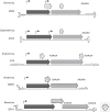
Figure 1.
Figure 1.
Genetic strategies of representative viruses from genera in the subfamily Parvovirinae. Genomes from the type species of each genus are denoted as a single line terminating in hairpin structures. The hairpins, drawn to represent their predicted structures, are scaled about 20× with respect to the rest of the genome. Open reading frames are represented by arrowed boxes, shaded dark gray for the major SF3 domain-containing replication initiator protein, light gray for the structural proteins of the capsid, and cross-hatched for sequences unique to the ancillary nonstructural proteins. Ancillary proteins for MVM are NS2 (left) and SAT (right) (Zádori et al. 2005; Ruiz et al. 2011), AAP for AAV2 (Sonntag et al. 2011), 7.5 kDa (left) and 11 kDa (right) for B19 (Zhi et al. 2006), NS2 for AMDV (Oleksiewicz et al. 1996), and NP1 for BPV (Chen et al. 1986). Transcriptional promoters are indicated by solid arrows and polyadenylation sites by the AAAAA sequence block.
Figure 2.
Figure 2.
“Two portal” model for genome packaging and uncoating. (Top left) A newly displaced single-stranded progeny genome is depicted 3′-to-5′, left-to-right, with a sphere denoting a covalently bound NS1 molecule. Beneath this, the DNA is shown being introduced into a preassembled capsid in a 3′-to-5′ direction, likely via the helicase activity of an NS1 peptide oligomer (depicted as a hexameric structure, by analogy with AAV) (Yoon-Robarts et al. 2004), with the 3′ end of the entering genome lodging near the capsid shell, proximal to its destined exit pore. The rest of the strand is then pumped into the particle under increasing pressure, adopting a topology that will allow it to be subsequently unraveled in a 3′-to-5′ direction. NS1 is removed from the 5′ end of the DNA during cell entry. Ca2+/Mg2+ ions in the virion are required to stabilize the pressurized structure, but when these are depleted in vitro, perhaps mimicking a specific host factor trigger in vivo, the 3′ end of the DNA, followed by the coding sequences, is ejected. This uncoated DNA can support complementary strand synthesis, while the 5′ end of the negative strand genome remains threaded with the capsid.
Figure 3.
Figure 3.
Progressive development of viral replication centers. (AF) A classification scheme, with classes 0–IV, shows the progressive changes in NS1 distribution that emerge with time after synchronized A9 cells infected with wild-type MVMp are released into S phase. Classes 0 and I were first observed 3 h into S phase, class II at 6 h, and classes III and IV at 12 h, although the number of cells showing the latter class morphologies progressively increased with time (quantified in F, where the distribution of NS1 morphology classes is plotted against time in seconds). (GI) Accumulation of MDC1 DDR foci around APAR bodies (stained for NS1) in MVM-infected wild-type mouse embryonic fibroblasts. (From Ruiz et al. 2011; reproduced, with permission, from Elsevier © 2011.)

References

    1. Adeyemi RO, Landry S, Davis ME, Weitzman MD, Pintel DJ 2010. Parvovirus minute virus of mice induces a DNA damage response that facilitates viral replication. PLoS Pathog 6: e1001141. - PMC - PubMed
    1. Allander T, Tammi MT, Eriksson M, Bjerkner A, Tiveljung-Lindell A, Andersson B 2005. Cloning of a human parvovirus by molecular screening of respiratory tract samples. Proc Natl Acad Sci 102: 12891–12896 - PMC - PubMed
    1. Bashir T, Rommelaere J, Cziepluch C 2001. In vivo accumulation of cyclin A and cellular replication factors in autonomous parvovirus minute virus of mice-associated replication bodies. J Virol 75: 4394–4398 - PMC - PubMed
    1. Cervelli T, Palacios JA, Zentilin L, Mano M, Schwartz RA, Weitzman MD, Giacca M 2008. Processing of recombinant AAV genomes occurs in specific nuclear structures that overlap with foci of DNA-damage-response proteins. J Cell Science 121: 349–357 - PubMed
    1. Chen KC, Shull BC, Moses EA, Lederman M, Stout ER, Bates RC 1986. Complete nucleotide sequence and genome organization of bovine parvovirus. J Virol 60: 1085–1097 - PMC - PubMed

Publication types