. 2013 Feb 1;140(3):530-40.
doi: 10.1242/dev.088583.

Cyclin E controls Drosophila female germline stem cell maintenance independently of its role in proliferation by modulating responsiveness to niche signals

Affiliations

Cyclin E controls Drosophila female germline stem cell maintenance independently of its role in proliferation by modulating responsiveness to niche signals

Elizabeth T Ables et al. Development. .

Abstract

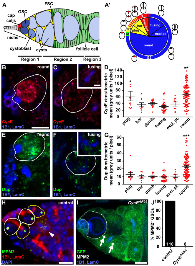
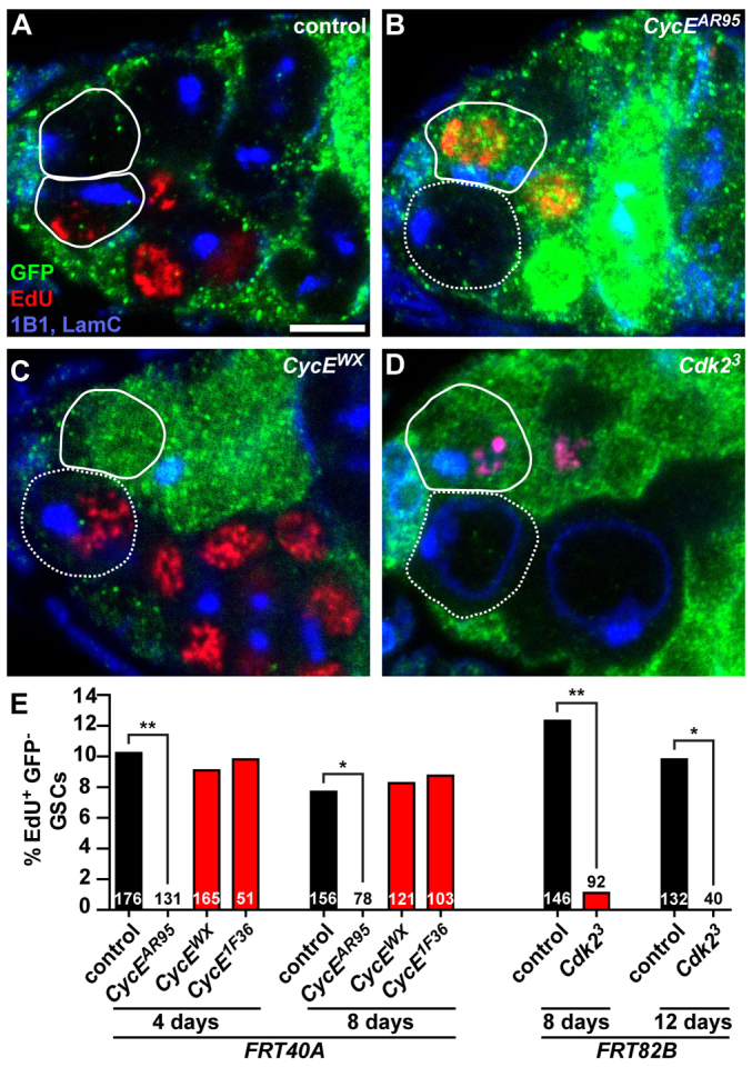
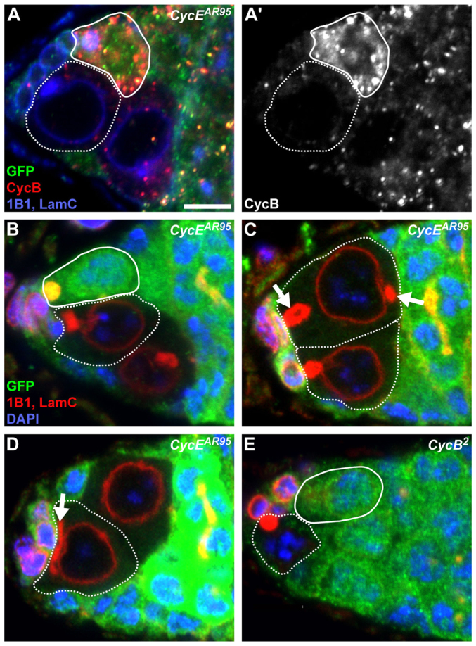
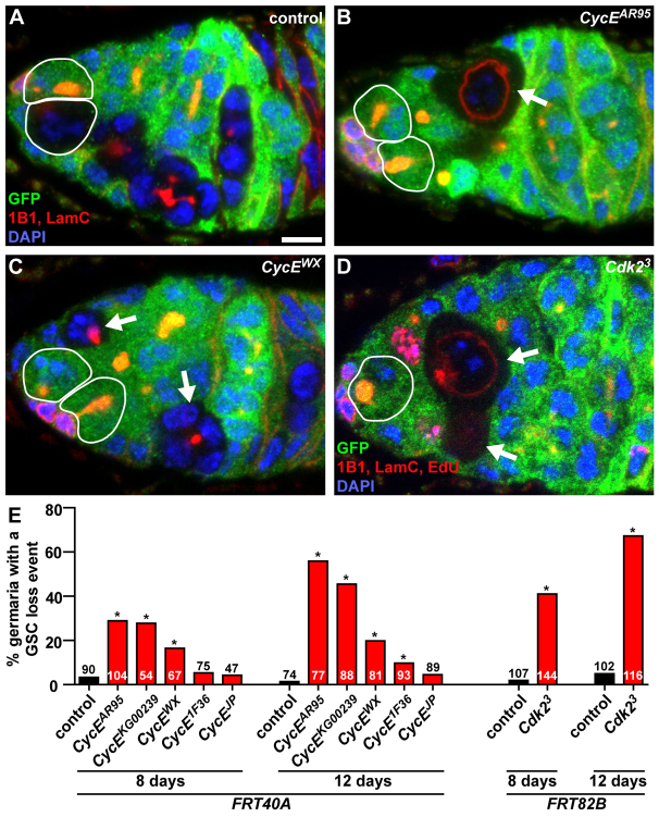
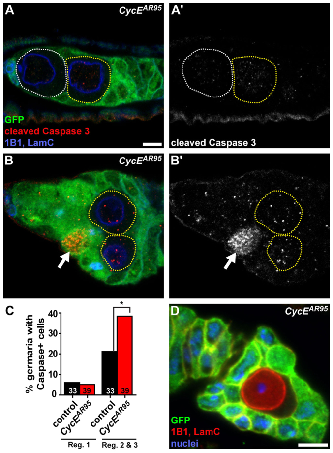
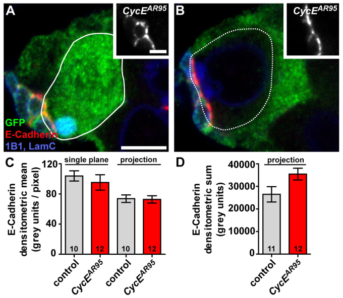
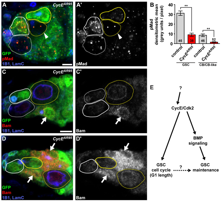
Stem cells must proliferate while maintaining 'stemness'; however, much remains to be learned about how factors that control the division of stem cells influence their identity. Multiple stem cell types display cell cycles with short G1 phases, thought to minimize susceptibility to differentiation factors. Drosophila female germline stem cells (GSCs) have short G1 and long G2 phases, and diet-dependent systemic factors often modulate G2. We previously observed that Cyclin E (CycE), a known G1/S regulator, is atypically expressed in GSCs during G2/M; however, it remained unclear whether CycE has cell cycle-independent roles in GSCs or whether it acts exclusively by modulating the cell cycle. In this study, we detected CycE activity during G2/M, reflecting its altered expression pattern, and showed that CycE and its canonical partner, Cyclin-dependent kinase 2 (Cdk2), are required not only for GSC proliferation, but also for GSC maintenance. In genetic mosaics, CycE- and Cdk2-deficient GSCs are rapidly lost from the niche, remain arrested in a G1-like state, and undergo excessive growth and incomplete differentiation. However, we found that CycE controls GSC maintenance independently of its role in the cell cycle; GSCs harboring specific hypomorphic CycE mutations are not efficiently maintained despite normal proliferation rates. Finally, CycE-deficient GSCs have an impaired response to niche bone morphogenetic protein signals that are required for GSC self-renewal, suggesting that CycE modulates niche-GSC communication. Taken together, these results show unequivocally that the roles of CycE/Cdk2 in GSC division cycle regulation and GSC maintenance are separable, and thus potentially involve distinct sets of phosphorylation targets.


Figures

Fig. 1.
Fig. 1.
CycE expression peaks during G2, leading to an atypical CycE activity pattern in GSCs. (A) Drosophila germarium. GSCs in a niche composed of terminal filament (grey), cap cells and a subset of escort cells (yellow) give rise to cystoblasts that form 16-cell cysts. (A′) Distribution of GSCs displaying specific fusome morphologies relative to cell cycle phases (supplementary material Fig. S1, Table S1). (B-G) Expression of CycE (B-D) and Dup (E-G) in GSCs. (B,C,E,F) CycE (red; B,C) or Dup (green; E,F) expression in GSCs (outlined) with ‘round’ (B,E) or ‘fusing’ (C,F) fusomes; insets show GSC fusomes, visible in adjacent optical slices. (D,G) Average CycE (D) or Dup (G) fluorescence intensity in GSCs according to fusome morphology. Bars represent s.e.m. *P<0.05, compared with ‘fusing’; **P<0.05, compared with ‘bar’, ‘dumbbell’, ‘fusing’ and ‘exclamation point’; ***P<0.05, compared with all other fusome morphologies. (H) Maximum intensity projection (6 μm thickness) showing MPM2 (green) labeling the histone locus body in GSCs (white outlines) and nascent cystoblasts (yellow outlines). In mitotically dividing cysts (e.g. eight-cell cystocyte indicated by an arrowhead), MPM2 expression oscillates, as described previously (Narbonne-Reveau and Lilly, 2009). (I) MPM2 (white) labeling the histone locus body (arrows) in wild-type germline cells (marked by GFP; green), but absent from a GFP-negative CycE-null GSC (circled). (B,C,E,F,I) 1B1, fusomes (blue); LamC, cap cell nuclear envelopes (blue). LamC is also prominent in CycE mutant germ cells. Scale bars: 5 μm in all main panels or 2.5 μm in insets in C,F. (J) Quantification of MPM2-positive wild-type versus CycE-deficient GSCs. Numbers in bars represent the number of GSCs analyzed.
Fig. 2.
Fig. 2.
CycE and Cdk2 are required for GSC proliferation. (A-D) Mosaic germaria showing control (A), null CycEAR95 (B), hypomorphic CycEWX (C) and null Cdk23 (D) GFP-negative clones. GFP (green), wild-type control cells; EdU (red), cells in S phase; 1B1 (blue), fusomes; LamC (blue), cap cell nuclear envelopes. Scale bar: 5 μm. (E) Percentage of GFP-negative GSCs that are positive for EdU in mosaic germaria at 4 or 8 days after clone induction. Numbers in the bars represent the number of GFP-negative GSCs analyzed. *P≤0.01; **P≤0.001.
Fig. 3.
Fig. 3.
GSCs lacking CycE arrest in a G1-like state showing a phenotype distinct from that of CycB-deficient GSCs. (A,A′) Mosaic germarium showing a GFP-negative CycEAR95 clone. CycB (red) is shown in greyscale in A′. GFP (green), wild-type control cells; 1B1 (blue), fusomes; LamC (blue), cap cell nuclear envelopes. (B-E) CycEAR95 (B-D) or CycB2 (E) mosaic germaria showing GFP-negative clones. Arrows indicate unusual fusome morphology (see supplementary material Fig. S1 for comparison). GFP (green), wild-type control cells; 1B1 (red), fusomes; LamC (red), cap cell nuclear envelopes; DAPI (blue), nuclei. Solid outline indicates wild-type GSCs; dashed outlines indicate mutant GSCs. Scale bar: 5 μm.
Fig. 4.
Fig. 4.
CycE and Cdk2 are required for GSC maintenance; this role is at least in part genetically separable from cell cycle regulation. (A-D) Mosaic germaria showing control (A), null CycEAR95 (B), hypomorphic CycEWX (C) and null Cdk23 (D) GFP-negative clones. GFP (green), wild-type control cells; 1B1 (red), fusomes; LamC (red), cap cell nuclear envelopes; DAPI (blue), nuclei. GSCs are outlined. Arrows indicate GSC loss events. Scale bar: 5 μm. (E) Percentage of germline-mosaic germaria with a GSC loss event 8 or 12 days after clone induction. Numbers in the bars represent the number of germline-mosaic germaria analyzed. *P<0.0001.
Fig. 5.
Fig. 5.
CycE mutant GSCs are not lost through apoptosis. (A-B′) Mosaic germaria showing GFP-negative CycEAR95 clones. GFP (green), wild-type control cells; cleaved Caspase 3 (red) (greyscale images in A′,B′), early apoptosis marker; 1B1 (blue), fusomes; LamC (blue), cap cell nuclear envelopes. CycEAR95 GSCs are outlined in white; CycEAR95 cystoblast-like cells are outlined in yellow. Arrows indicate a GFP-positive wild-type germ cell positive for cleaved Caspase 3. (C) Percentage of germline-mosaic germaria with at least one Caspase-positive cell in region 1 (which contains GSCs and cystoblasts) or regions 2 and 3 (which normally contain more differentiated cysts) in ‘mock’ control (black) or CycEAR95 (red) mosaic germaria. Numbers in bars represent number of germline-mosaic germaria analyzed. *P<0.01. (D) Single GFP-negative CycEAR95 germ cell enveloped by follicle cells in mosaic ovariole. GFP (green), wild-type control cells; 1B1 (red), follicle cell membranes. LamC (red) stains germ cell nuclear envelope; DAPI (blue), nuclei. Scale bars: 5 μm.
Fig. 6.
Fig. 6.
CycE AR95 GSCs have normal levels of E-cadherin at the GSC-cap cell interface. (A,B) Anterior region of a mosaic germarium showing a GFP-positive wild-type GSC (A) and a GFP-negative CycEAR95 GSC (B). GFP (green), wild-type control cells; E-cadherin (red; greyscale images shown in insets); 1B1 (blue), fusomes; LamC (blue) cap cell nuclear envelopes. GSCs are outlined. Scale bars: 5 μm (or 2.5 μm in insets). (C,D) E-cadherin levels at the GSC-cap cell junction, measured by densitometric mean (C) or sum (D) of pixel intensity in single optical slices or maximum intensity projections in CycEAR95 or adjacent wild-type GSCs. Numbers in the bars represent number of GSCs measured. Bars represent s.e.m.
Fig. 7.
Fig. 7.
CycE AR95 GSCs have reduced BMP signaling, but do not fully differentiate. (A,A′) CycEAR95 mosaic germaria showing GFP-negative clones. pMad (red; greyscale image in A′) is a BMP signaling marker. Arrowheads indicate a CycEAR95 cystoblast-like cell. (B) Average level of pMad expression in control or CycEAR95 GSCs in mosaic germaria. Numbers in the bars represent the number of GSCs analyzed. Bars represent s.e.m. **P<0.01. (C-D′) CycEAR95 mosaic germaria. Bam (red; greyscale image in C′,D′) is an early differentiation marker. Arrows indicate GFP-positive wild-type germline cysts that express high levels of Bam. In A,C,D, GFP (green) indicates control cells, 1B1 (blue) indicates fusomes, and LamC (blue) indicates cap cell nuclear envelopes. Control wild-type GSCs have solid white outlines; CycEAR95 GSCs are indicated by dashed white outlines; control cystoblasts are indicated by solid yellow outlines; CycEAR95 cystoblast-like cells are indicated by dashed yellow outlines. (E) Model for genetically separable roles of CycE/Cdk2 in regulating the female GSC cell cycle and maintenance. CycE function promotes GSC responsiveness to niche BMP signals; in addition, high CycE expression during G2 and M probably results in rapid entry into S following M phase, which limitis excessive growth during G1. An indirect contribution of CycE/Cdk2 to GSC function through its effect on the cell cycle is also possible. It remains unknown what mechanisms lead to the atypical CycE pattern in GSCs. Scale bars: 5 μm.

References

    1. Ables E. T., Drummond-Barbosa D. (2010). The steroid hormone ecdysone functions with intrinsic chromatin remodeling factors to control female germline stem cells in Drosophila. Cell Stem Cell 7, 581-592 - PMC - PubMed
    1. Ables E. T., Laws K. M., Drummond-Barbosa D. (2012). Control of adult stem cells in vivo by a dynamic physiological environment: diet-dependent systemic factors in Drosophila and beyond. WIREs Dev Biol 1, 657-674 - PMC - PubMed
    1. Ashburner M., Drysdale R. (1994). FlyBase – the Drosophila genetic database. Development 120, 2077-2079 - PubMed
    1. Ballabeni A., Park I. H., Zhao R., Wang W., Lerou P. H., Daley G. Q., Kirschner M. W. (2011). Cell cycle adaptations of embryonic stem cells. Proc. Natl. Acad. Sci. USA 108, 19252-19257 - PMC - PubMed
    1. Berger C., Pallavi S. K., Prasad M., Shashidhara L. S., Technau G. M. (2005). A critical role for cyclin E in cell fate determination in the central nervous system of Drosophila melanogaster. Nat. Cell Biol. 7, 56-62 - PubMed

Publication types

MeSH terms