. 2013 Feb 1;140(3):583-93.
doi: 10.1242/dev.085118.

Lrp4 and Wise interplay controls the formation and patterning of mammary and other skin appendage placodes by modulating Wnt signaling

Affiliations

Lrp4 and Wise interplay controls the formation and patterning of mammary and other skin appendage placodes by modulating Wnt signaling

Youngwook Ahn et al. Development. .

Abstract

The future site of skin appendage development is marked by a placode during embryogenesis. Although Wnt/β-catenin signaling is known to be essential for skin appendage development, it is unclear which cellular processes are controlled by the signaling and how the precise level of the signaling activity is achieved during placode formation. We have investigated roles for Lrp4 and its potential ligand Wise (Sostdc1) in mammary and other skin appendage placodes. Lrp4 mutant mice displayed a delay in placode initiation and changes in distribution and number of mammary precursor cells leading to abnormal morphology, number and position of mammary placodes. These Lrp4 mammary defects, as well as limb defects, were associated with elevated Wnt/β-catenin signaling and were rescued by reducing the dose of the Wnt co-receptor genes Lrp5 and Lrp6, or by inactivating the gene encoding β-catenin. Wise-null mice phenocopied a subset of the Lrp4 mammary defects and Wise overexpression reduced the number of mammary precursor cells. Genetic epistasis analyses suggest that Wise requires Lrp4 to exert its function and that, together, they have a role in limiting mammary fate, but Lrp4 has an early Wise-independent role in facilitating placode formation. Lrp4 and Wise mutants also share defects in vibrissa and hair follicle development, suggesting that the roles played by Lrp4 and Wise are common to skin appendages. Our study presents genetic evidence for interplay between Lrp4 and Wise in inhibiting Wnt/β-catenin signaling and provides an insight into how modulation of Wnt/β-catenin signaling controls cellular processes important for skin placode formation.


Figures

Fig. 1.
Fig. 1.
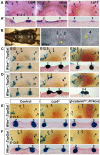
Abnormal mammary development in Lrp4 mutant mice. (A) Five pairs of nipples in a pregnant control female. (B) Lrp4 mutant female displays ectopic nipples (arrowheads) and fusion of nipples 2 and 3. (C) The appearance of mammary placodes during embryogenesis (top) and distribution of mammary epithelial cells around placodes 2 and 3 (bottom). (D,E) TopGal-expressing epithelial cells are gradually restricted to placodes in controls. In Lrp4 mutants, delayed placode formation (E12.0) is followed by ectopic placodes (arrows) and fusion of placodes 2 and 3. Higher magnification images (E12.0) and histological sections (E12.5 and E14.5) of the placode 2/3 area are shown below.
Fig. 2.
Fig. 2.
Increased number of mammary epithelial cells in Lrp4 mutants. (A,B) Confocal images of the placode 2/3 region from TCF/LEF:H2B-GFP embryos. (C) Relative number of GFP-positive cells as marked by a red dot in A,B. Data are mean±s.d. (D-G) BrdU staining is reduced in the interplacodal region (arrow) in Lrp4 mutants. (H,I) Caspase 3 staining. (J,K) E-cadherin staining. Note that placode 2 is out of the focal plane in F and J.
Fig. 3.
Fig. 3.
Genetic interaction of Lrp4 with Lrp5 and Lrp6. TopGal expression at E13.5. A low level of broad β-galactosidase activity is detectable from the Lrp6 mutant allele. (A-B′) Reduced dosages of Lrp5 and Lrp6 rescue the limb (A) and mammary (B,B′) defects of Lrp4 mutants. A proximal (left, dorsal to the right) and a dorsal (right) view of a forelimb bud are shown with anterior to the top (A). (C,C′) Lrp4 and Lrp6 compensate for loss of each other in limbs. Note that hindlimb defects of Lrp6-/- mice were rescued by inactivation of Lrp4, but other defects such as loss of tail remain the same (arrows). (D) Separation of mammary bud #2 and 3 by reduced dosages of Lrp5 and Lrp6 in Lrp4 mutants.
Fig. 4.
Fig. 4.
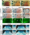
Lrp4 facilitates placode initiation and controls the number of mammary epithelial cells via inhibition of Wnt/β-catenin signaling. (A-C′) Reduced dose of Lrp5 and Lrp6 restores normal timing of placode initiation and reduces ectopic TopGal-expressing cells in Lrp4 mutants. (D-F′) Detection of Cre activity from K14cre transgene. (G-J′) Conditional inactivation of Ctnnb1 in control mice results in smaller mammary buds. In Lrp4 mutants, inactivation of Ctnnb1 results in separated, but much smaller, buds.
Fig. 5.
Fig. 5.
Lrp4 is required for development of other skin appendages. (A) Delayed formation of the primary hair follicles in Lrp4 mutants, as evidenced by lack of Wnt10b expression. (B-E′) Expression of the Lrp4-lacZ BAC reporter line in the primary hair follicles and mammary buds (1-5) at E13.5-E14.5. Focalized reporter expression is normally observed in mature hair placodes of back skin at E14.5. In Lrp4 mutants, Lrp4-lacZ expression is spread along the mammary line (arrow) with no sign of hair placodes at E13.5 (C) and hair follicle morphogenesis is delayed (E,E′). (F-G′) Abnormal patterning of interramal vibrissal follicles in Lrp4 mutants. Frontal sections (F′-G′) were obtained along the broken line. (H,I) Lrp4 mutants display supernumerary vibrissal follicles in the submental (rectangle), postoral (circle) and interramal (oval) regions.
Fig. 6.
Fig. 6.
Wise controls patterning of mammary placodes via the Wnt/β-catenin pathway. (A,A′) Whole-mount in situ hybridization (A) and cross-section across mammary placodes 2 and 3 (A′). (B) Wise-null females display abnormal spacing between nipples and supernumerary nipples (arrowheads). (C,D) In mutants (D), abnormal size and morphology of the placodes is apparent by E12.5. The distance between placodes 2 and 3 is reduced, often leading to fusion at later stages. TopGal-expressing cells are ectopically observed in the interplacodal regions. (E,F) Loss of Lrp5 or epithelial inactivation of Ctnnb1 restores normal spacing between mammary buds 2 and 3 in Wise-null mice.
Fig. 7.
Fig. 7.
Wise overexpression disrupts mammary development. (A) K14-tTA and tetO-Wise constructs. (B-C′) With a strong driver, Wise overexpression disrupts limb development (arrow) and results in smaller mammary placodes. (D-G′) Wise-null mammary defects are rescued by a moderate level of Wise expression in the ectoderm. (H) TCF-tTA construct. (I-K′) TCF-tTA;tetO-Wise mice display limb and mammary defects. TopGal (B-G′,I-J′) and eGFP (K,K′).
Fig. 8.
Fig. 8.
Genetic interaction between Lrp4 and Wise. (A-C) TopGal expression in Lrp4;Wise double mutant mice. Transheterozygotes display normal mammary patterning, and inactivation of Wise does not exacerbate Lrp4 mutant defects such as fusion of buds 2 and 3, and ectopic buds (arrows). (D-I′) Wise overexpression, as evidenced by eGFP expression (G′-I′), fails to rescue the mammary (D,E,G,H) and forelimb (F,I) defects of Lrp4 mutants, as shown by no significant change in TopGal expression. Arrows indicate ectopic mammary placodes. (J-M′) Wise overexpression causes reduction in the number of vibrissal follicles (J,L) and taste papilla (J′,L′), but not in Lrp4 mutants (K,M,K′,M′). All at E14.5 except F,I,I′, which are at E13.5.
Fig. 9.
Fig. 9.
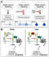
Model for function of Lrp4 and Wise in mammary development. (A) Wnt/β-catenin signaling modulates multiple steps of placode development. Initially, Lrp4 facilitates placode initiation, and later Lrp4 and Wise together limit the number of mammary precursor cells by inhibition of Wnt/β-catenin signaling. (B) Early (left), Lrp4 functions in a Wise-independent manner, but later (right), Lrp4 and Wise act together to inhibit Wnt/β-catenin signaling.

References

    1. Ahn Y., Sanderson B. W., Klein O. D., Krumlauf R. (2010). Inhibition of Wnt signaling by Wise (Sostdc1) and negative feedback from Shh controls tooth number and patterning. Development 137, 3221-3231. - PMC - PubMed
    1. Balemans W., Ebeling M., Patel N., Van Hul E., Olson P., Dioszegi M., Lacza C., Wuyts W., Van Den Ende J., Willems P., et al. (2001). Increased bone density in sclerosteosis is due to the deficiency of a novel secreted protein (SOST). Hum. Mol. Genet. 10, 537-543. - PubMed
    1. Balinsky B. I. (1950). On the prenatal growth of the mammary gland rudiment in the mouse. J. Anat. 84, 227-235. - PMC - PubMed
    1. Boras-Granic K., Chang H., Grosschedl R., Hamel P. A. (2006). Lef1 is required for the transition of Wnt signaling from mesenchymal to epithelial cells in the mouse embryonic mammary gland. Dev. Biol. 295, 219-231. - PubMed
    1. Brault V., Moore R., Kutsch S., Ishibashi M., Rowitch D. H., McMahon A. P., Sommer L., Boussadia O., Kemler R. (2001). Inactivation of the beta-catenin gene by Wnt1-Cre-mediated deletion results in dramatic brain malformation and failure of craniofacial development. Development 128, 1253-1264. - PubMed

Publication types

MeSH terms

Substances

LinkOut - more resources