. 2013 Feb 1;140(3):639-48.
doi: 10.1242/dev.086702.

Sox17 haploinsufficiency results in perinatal biliary atresia and hepatitis in C57BL/6 background mice

Affiliations

Sox17 haploinsufficiency results in perinatal biliary atresia and hepatitis in C57BL/6 background mice

Mami Uemura et al. Development. .

Abstract

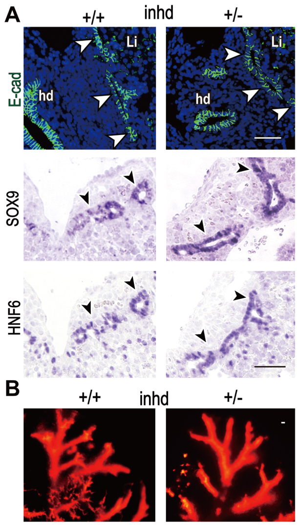
Congenital biliary atresia is an incurable disease of newborn infants, of unknown genetic causes, that results in congenital deformation of the gallbladder and biliary duct system. Here, we show that during mouse organogenesis, insufficient SOX17 expression in the gallbladder and bile duct epithelia results in congenital biliary atresia and subsequent acute 'embryonic hepatitis', leading to perinatal death in ~95% of the Sox17 heterozygote neonates in C57BL/6 (B6) background mice. During gallbladder and bile duct development, Sox17 was expressed at the distal edge of the gallbladder primordium. In the Sox17(+/-) B6 embryos, gallbladder epithelia were hypoplastic, and some were detached from the luminal wall, leading to bile duct stenosis or atresia. The shredding of the gallbladder epithelia is probably caused by cell-autonomous defects in proliferation and maintenance of the Sox17(+/-) gallbladder/bile duct epithelia. Our results suggest that Sox17 plays a dosage-dependent function in the morphogenesis and maturation of gallbladder and bile duct epithelia during the late-organogenic stages, highlighting a novel entry point to the understanding of the etiology and pathogenesis of human congenital biliary atresia.


Figures

Fig. 1.
Fig. 1.
Defective development of the liver and gallbladder/bile duct in Sox17 heterozygote embryos in B6 background mice. (A,B) Gross-anatomical (upper panels in A) and histological [PAS staining (middle) and HE staining (lower panels in A)] images showing peripheral degeneration (asterisks and insets in A) of the liver lobules in Sox17+/− embryos at 17.5 dpc. In B, the line graph indicates significant reduction of liver weight in Sox17+/− embryos at 16.5 and 17.5 dpc compared with wild-type littermates (**P<0.01). Numbers in parentheses indicate the total number of the embryos used in each group. Error bars represent s.e.m. (C) Whole-mount DBA staining shows hypoplastic gallbladder (gb, insets) and ectopic formation of extrahepatic ducts (white arrowheads) in Sox17+/− embryos during 13.5 to 17.5 dpc. cb, common bile duct; cd, cystic duct; du, duodenum; gb, gallbladder; hd, extrahepatic duct. Scale bar: 50 μm.
Fig. 2.
Fig. 2.
Hepatic inflammation with elevated bile acid levels and endoplasmic reticulum stress of hepatocytes in fetal Sox17+/− B6 livers. (A) Real-time RT-PCR analysis showing elevated expression levels of several inflammatory cytokine markers in Sox17+/− B6 livers at 17.0 dpc. The vertical axis represents fold changes in the expression level of each liver sample relative to those of wild-type livers (the mean value was set as 1). Filled (severe) and unfilled (mild) circles indicate Sox17+/− liver samples with and without any gross-anatomical lesions, respectively. The bars show mean values of all (severe and mild) samples, whereas asterisks on the bar indicate significant differences compared with wild-type livers (*P<0.05, **P<0.01). (B) Biochemical data showing significantly elevated levels of serum ALT, serum ALP and intrahepatic bile acids in Sox17+/− livers compared with wild-type livers. Error bars represent s.e.m. (C) PAS (red) and anti-GRP78 immunostaining (brown) of two consecutive sections, showing GRP78-positive signals in the hepatocytes around the degenerating peripheral region of Sox17+/− liver lobules (arrows). Asterisks indicate the PAS-negative region including the damaged hepatocytes. (D) Electron microscopic images showing enlargement of rough ER (arrows) in Sox17+/− hepatocytes. Each dashed rectangle indicates the area highly magnified in the adjacent panel. bi, bile canaliculus; bv, blood vessel; hc, hepatocyte, rb, red blood (hematopoietic) cells. Scale bars: 50 μm in upper panels in C; 5 μm in lower right inset of C and in D.
Fig. 3.
Fig. 3.
SOX17 is highly expressed in proliferating gallbladder epithelia, but not in hepatocytes during the late-organogenic stages. (A-F) Anti-SOX17 (purple in A and C, red fluorescence in E) and anti-PCNA (green fluorescence in E and F) immunostaining of wild-type embryos and GFP fluorescence images of Sox17GFP/+ embryos (green fluorescence in B and D) at 13.5 dpc (E) and 16.5 dpc (A-D,F) showing SOX17 expression sites in developing liver (A,B) and gallbladder/bile duct (C-F). In A and B, the two lower insets indicate higher-magnified images of the surface (upper) and proximal (lower) regions of the liver lobules surrounded by the broken rectangles in the top panels, respectively. In C and D, left panels show SOX17 expression in whole-mount images of the gallbladder/bile-duct, whereas right panels display the transverse section images corresponding to the plane of the dashed lines at the levels of extrahepatic duct, cystic duct and gallbladder. (E,F) Anti-SOX17 (red) and anti-PCNA (green) double-immunostaining of the sagittal section (E) and anti-PCNA staining of the transverse section (F) of the gallbladder/bile-duct (DAPI, blue). Asterisk indicates non-specific fluorescence. cd, cystic duct; hd, extrahepatic duct; gb, gallbladder; v, blood vessel. Scale bars: 50 μm.
Fig. 4.
Fig. 4.
Hypoplasia of Sox17+/− gallbladder epithelium during late-organogenic stages. (A,B) Light (A; HE staining) and electron (B) microscopic images of gallbladder epithelia (transverse sections at the levels of maximum diameter), showing hypoplasia of Sox17+/− gallbladders at 15.5 to 17.5 dpc. Gallbladder epithelial cells showed defective maturation (vertical bars indicate epithelial cell height in A and basal lamina thickness in B) in Sox17+/− embryos. Arrows in A and B indicate presumptive decidual cells within the gallbladder epithelia. (C) Anti-PCNA immunofluorescence images (left panels) and quantitative data (right graph) showing significant reductions in the PCNA-positive index (**P<0.01 at 15.5 and 16.5 dpc) in Sox17+/− gallbladder epithelia (n=10 embryos in each bar). Error bars represent s.e.m. gb, gallbladder; ge, gallbladder epithelial cells. Scale bars: 50 μm in A,C; 5 μm in B.
Fig. 5.
Fig. 5.
Epithelail cell deciduation in the gallbladder, and biliary obstruction in the cystic duct and extrahepatic duct regions of Sox17+/− embryos. (A-E) Light (HE, DBA-lectin, anti-HNF6 and anti-E-cadherin (E-cad) staining) and electron microscopic images showing epithelial cell deciduation in the gallbladder (A-C) and luminal sloughed cells/cell debris in the cystic and extrahepatic ducts (arrowheads in D,E). Sloughed epithelial cells are positive for DBA lectin staining and anti-HNF6/E-cad immunostaining (arrowheads in A,B). In the gallbladder/cystic duct regions, luminal decidual cells can occupy the luminal space (right panel in B; and C). In the center of the obstruction region, the decidual cells undergo necrosis (asterisk in C2), whereas several outer decidual cells are tightly connected to the luminal cell surface of the bile duct epithelia (C3). In the cystic duct and extrahepatic duct regions at 15.5-17.5 dpc, the decidual cells are frequently found in the lumen (arrowheads in D and left panels of E), resulting in biliary atresia in the extrahepatic ducts (arrowheads in two right panels of E; the arrows indicate intrahepatic bile ducts). cd, cystic duct; dc, decidual cells in the lumen; gb, gallbladder; hd, extrahepatic duct; Li, liver parenchyma. Scale bars: 50 μm (except for 5 μm in C2,C3).
Fig. 6.
Fig. 6.
No appreciable defects in the formation and branching of intrahepatic biliary trees in fetal Sox17+/− livers. (A,B) Anti-E-cad (green fluorescence), SOX9 and HNF6 (purple) immunostaining (A) and a tracer experiment by injecting fluorescent ink into the gallbladder under high pressure (red fluorescence; B) revealed proper formation of the intrahepatic duct tree (arrowheads), even in Sox17+/− livers with gross-anatomical hepatic lesions at 17.0 dpc. hd, extrahepatic duct; inhd, intrahepatic bile duct; Li, liver parenchyma. Scale bars: 50 μm.
Fig. 7.
Fig. 7.
Reduced elongation, non-polarized proliferation, and shredding of epithelial cells of Sox17+/− gallbladder primordium in vitro. (A,B) Schematic (A) and phase-contrast images of the timecourse (B) of a 3-day organ culture of gallbladder primordium initiated at 13.5 dpc. The dashed arrows show the sectioning planes corresponding to C, D and E. (C-E) DBA-stained whole-mount images (horizontal view; C); anti-PCNA-, anti-laminin-, and DBA-stained section images (horizontal section; C), HE- and anti-E-cad/anti-ZO1-stained images (transverse sections; D); and anti-integrin β1-stained images (transverse sections; E) of Sox17+/− and wild-type explants initiated at 13.5 dpc (3-day culture). The wild-type gallbladder explants showed polarized elongation of the ductural structure (B), polarized distribution of PCNA-positive epithelial cells along the distal-proximal axis (white unbroken arrow: PCNA-negative proximal region in C), and epithelial fold formation (D) in the developing gallbladder primordium in vitro. By contrast, Sox17+/− explants displayed a non-polarized balloon-like structure surrounded by a single thin epithelial cell layer (B-E), non-polarized distribution of PCNA-positive epithelial cells (C), a partial lack of anti-lamimin signals with the decidual epithelial cells (dashed arrow and filled arrowhead in C), and reduced anti-integrin β1 signals at the basal surface of the epithelial cells (E). The bottom panel in C show the Sox17+/− decidual epithelial cells indicated by arrowheads in the upper panels. Scale bar: 50 μm.

References

    1. Artus J., Piliszek A., Hadjantonakis A. K. (2011). The primitive endoderm lineage of the mouse blastocyst: sequential transcription factor activation and regulation of differentiation by Sox17. Dev. Biol. 350, 393-404 - PMC - PubMed
    1. Bamford R. N., Roessler E., Burdine R. D., Saplako lu U., dela Cruz J., Splitt M., Goodship J. A., Towbin J., Bowers P., Ferrero G. B., et al. (2000). Loss-of-function mutations in the EGF-CFC gene CFC1 are associated with human left-right laterality defects. Nat. Genet. 26, 365-369 - PubMed
    1. Bernstein H., Payne C. M., Bernstein C., Schneider J., Beard S. E., Crowley C. L. (1999). Activation of the promoters of genes associated with DNA damage, oxidative stress, ER stress and protein malfolding by the bile salt, deoxycholate. Toxicol. Lett. 108, 37-46 - PubMed
    1. Bochkis I. M., Rubins N. E., White P., Furth E. E., Friedman J. R., Kaestner K. H. (2008). Hepatocyte-specific ablation of Foxa2 alters bile acid homeostasis and results in endoplasmic reticulum stress. Nat. Med. 14, 828-836 - PMC - PubMed
    1. Cardinale V., Wang Y., Carpino G., Cui C. B., Gatto M., Rossi M., Berloco P. B., Cantafora A., Wauthier E., Furth M. E., et al. (2011). Multipotent stem/progenitor cells in human biliary tree give rise to hepatocytes, cholangiocytes, and pancreatic islets. Hepatology 54, 2159-2172 - PubMed

Publication types

MeSH terms