. 2013 Feb 1;140(3):660-6.
doi: 10.1242/dev.088526.

In vivo monitoring of cardiomyocyte proliferation to identify chemical modifiers of heart regeneration

Affiliations

In vivo monitoring of cardiomyocyte proliferation to identify chemical modifiers of heart regeneration

Wen-Yee Choi et al. Development. .

Abstract

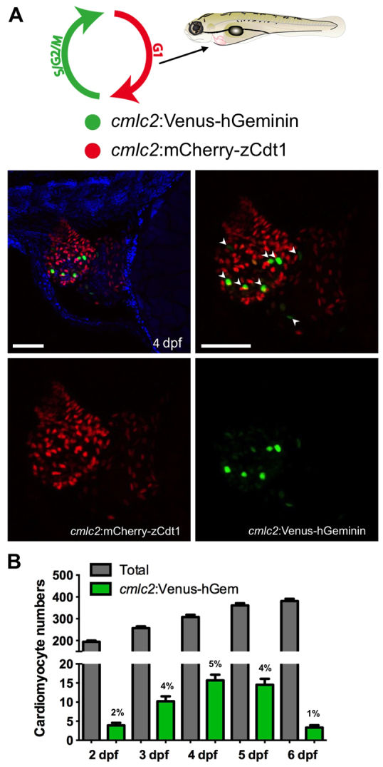
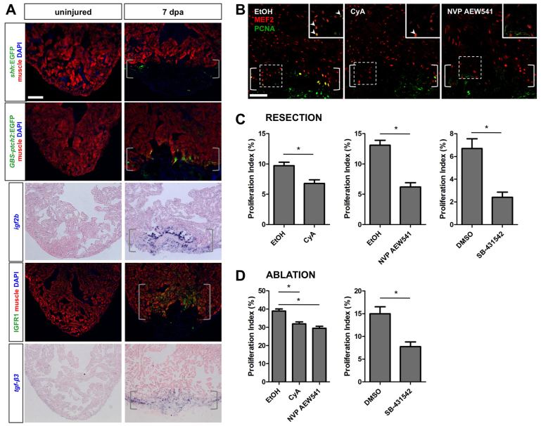
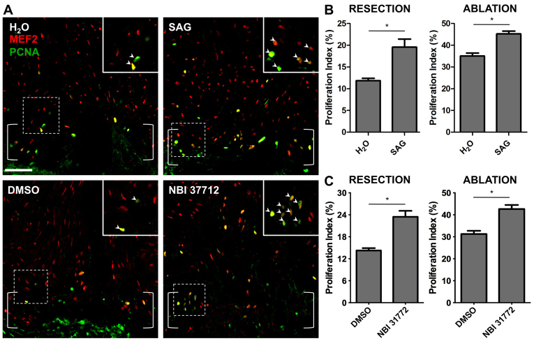
Adult mammalian cardiomyocytes have little capacity to proliferate in response to injury, a deficiency that underlies the poor regenerative ability of human hearts after myocardial infarction. By contrast, zebrafish regenerate heart muscle after trauma by inducing proliferation of spared cardiomyocytes, providing a model for identifying manipulations that block or enhance these events. Although direct genetic or chemical screens of heart regeneration in adult zebrafish present several challenges, zebrafish embryos are ideal for high-throughput screening. Here, to visualize cardiomyocyte proliferation events in live zebrafish embryos, we generated transgenic zebrafish lines that employ fluorescent ubiquitylation-based cell cycle indicator (FUCCI) technology. We then performed a chemical screen and identified several small molecules that increase or reduce cardiomyocyte proliferation during heart development. These compounds act via Hedgehog, Insulin-like growth factor or Transforming growth factor β signaling pathways. Direct examination of heart regeneration after mechanical or genetic ablation injuries indicated that these pathways are activated in regenerating cardiomyocytes and that they can be pharmacologically manipulated to inhibit or enhance cardiomyocyte proliferation during adult heart regeneration. Our findings describe a new screening system that identifies molecules and pathways with the potential to modify heart regeneration.


Figures

Fig. 1.
Fig. 1.
FUCCI zebrafish for visualizing cardiomyocyte proliferation. (A) Representative maximum intensity projection of the heart of a 4 dpf cmlc2:FUCCI transgenic larva, visualizing non-proliferating (cmlc2:mCherry-zCdt1, red) and proliferating (cmlc2:Venus-hGeminin, green) cardiomyocytes, as depicted in the schematic at the top. Cardiomyocytes with only the Venus-hGeminin signal (arrowheads) were considered to be proliferating. DAPI, blue. Scale bars: 50 μm. (B) Time course of total (red+ and green+) and proliferating (green+) cardiomyocytes from 2 to 6 dpf. Percentages of proliferating cardiomyocytes are indicated above bars. Data are represented as mean±s.e.m. n=13-24 embryos per stage.
Fig. 2.
Fig. 2.
Hedgehog and Igf signaling promote embryonic cardiomyocyte proliferation in zebrafish. (A) Small molecule screen to identify signaling pathways that affect cardiomyocyte (CM) proliferation between 3 and 4 dpf. Green bars, drugs that were identified as potential enhancers of proliferation; red bars, drug that was identified as a potential inhibitor. Data are represented as mean±s.e.m. n=8-12 embryos per condition. In the schematic, green and red circles represent proliferating and non-proliferating cells, respectively. (B) Representative maximum intensity projections of 4 dpf cmlc2:FUCCI embryos treated with Smoothened agonist (SAG) or the Igf agonist NBI-31772. Scale bars: 50 μm. Anterior is to the left in images. (C) Effects of treatments with SAG (5 μM), CyA (5 μM), NBI-31772 (2.5 μM), NVP AEW541 (5 μM) and SB-431542 (5 μM) on cardiomyocyte proliferation signals. n=30-48, mean±s.e.m. *P<0.005, Student's t-test. Experiments for an individual graph were performed on a combined pool of embryos. Proliferation indices could vary between pools used for each individual graph. (D) Changes in proliferation were reflected by analogous changes in the total number of cardiomyocytes in cmlc2:nucDsRed2 embryos at 4 dpf. n=23-35, mean±s.e.m. *P<0.05, Student's t-test.
Fig. 3.
Fig. 3.
Hedgehog, Igf and Tgfβ signaling are required for myocardial regeneration in zebrafish. (A) The Shh ligand shha (transgenic reporter) and regulator/response gene ptch2 (transgenic reporter), Igf ligand igf2b (in situ hybridization) and receptor Igfr1 (immunofluorescence), and Tgfβ ligand tgfb3 (in situ hybridization) each showed increased expression in the wound area by 7 dpa. Brackets indicate injury site. Scale bar: 100 μm. (B) Treatment with CyA (10 μM) or NVP AEW541 (2 μM) from 6 to 7 dpa decreased cardiomyocyte proliferation. Mef2, red; PCNA, green. Brackets indicate injury site. Insets: High magnifications of the boxed areas. Arrowheads indicate proliferating cardiomyocytes. Scale bar: 50 μm. (C) Quantification of the effects of CyA (10 μM), NVP AEW541 (2 μM) and SB-431542 (10 μM) on cardiomyocyte proliferation after resection injury. Fish were treated from 6 to 7 dpa. n=9-15, mean±s.e.m. *P<0.01, Student's t-test. (D) Quantification of the effects of CyA, NVP AEW541 and SB-431542 on proliferation after genetic cardiomyocyte ablation. Fish were treated from 6 to 7 dpa. n=12-13, mean±s.e.m. *P<0.001, Student's t-test. Data in each graph were obtained from different clutches of fish.
Fig. 4.
Fig. 4.
Hedgehog or Igf pathway activation increases cardiomyocyte proliferation during regeneration in zebrafish. (A) Treatment with SAG (2.5 μM) or NBI-31772 (10 μM) from 6 to 7 dpa increased cardiomyocyte proliferation. Mef2, red; PCNA, green. Brackets indicate injury site. Insets: High magnification of the boxed areas. Arrowheads indicate proliferating cardiomyocytes. Scale bar: 50 μm. (B) Quantification of cardiomyocyte proliferation following treatment with SAG (2.5 μM) after resection (left) or ablation (right) injury models. Fish were treated from 6 to 7 dpa. n=9-15, mean±s.e.m. *P<0.01, Student's t-test. (C) Quantification of cardiomyocyte proliferation following treatment with NBI-31772 after resection (10 μM) or ablation (5 μM) injury models. n=11-17, mean±s.e.m. *P<0.05, Student's t-test.

References

    1. Bergmann O., Bhardwaj R. D., Bernard S., Zdunek S., Barnabé-Heider F., Walsh S., Zupicich J., Alkass K., Buchholz B. A., Druid H., et al. (2009). Evidence for cardiomyocyte renewal in humans. Science 324, 98-102 - PMC - PubMed
    1. Brade T., Kumar S., Cunningham T. J., Chatzi C., Zhao X., Cavallero S., Li P., Sucov H. M., Ruiz-Lozano P., Duester G. (2011). Retinoic acid stimulates myocardial expansion by induction of hepatic erythropoietin which activates epicardial Igf2. Development 138, 139-148 - PMC - PubMed
    1. Chablais F., Jaźwińska A. (2012). The regenerative capacity of the zebrafish heart is dependent on TGFβ signaling. Development 139, 1921-1930 - PubMed
    1. Chablais F., Veit J., Rainer G., Jaźwińska A. (2011). The zebrafish heart regenerates after cryoinjury-induced myocardial infarction. BMC Dev. Biol. 11, 21 - PMC - PubMed
    1. Chen J. K., Taipale J., Cooper M. K., Beachy P. A. (2002). Inhibition of Hedgehog signaling by direct binding of cyclopamine to Smoothened. Genes Dev. 16, 2743-2748 - PMC - PubMed

Publication types

MeSH terms

LinkOut - more resources