. 2013 Feb 1;190(3):1360-71.
doi: 10.4049/jimmunol.1200593. Epub 2013 Jan 4.

Heat shock protein 90 is critical for regulation of phenotype and functional activity of human T lymphocytes and NK cells

Affiliations

Heat shock protein 90 is critical for regulation of phenotype and functional activity of human T lymphocytes and NK cells

Jooeun Bae et al. J Immunol. .

Abstract

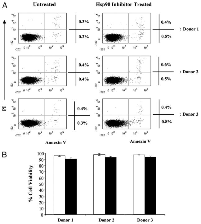
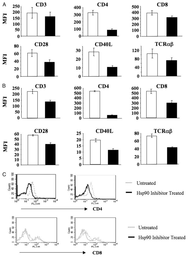
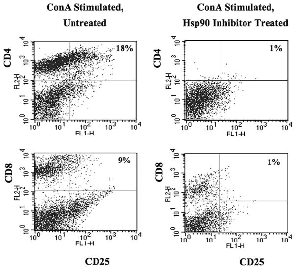
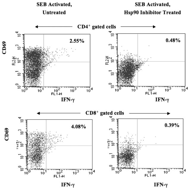
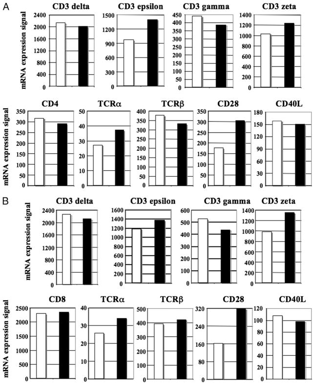
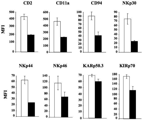
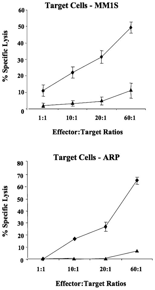
The 90-kDa heat shock protein (Hsp90) has become an important therapeutic target with ongoing evaluation in a number of malignancies. Although Hsp90 inhibitors have a high therapeutic index with limited effects on normal cells, they have been described to inhibit dendritic cell function. However, its effect on human immune effector cells may have significant clinical implications, but remains unexplored. In this study, we have evaluated the effects of Hsp90 inhibition on human T lymphocyte and NK cells, including their Ag expression, activation, proliferation, and functional activities. These studies demonstrate that Hsp90 inhibition irreversibly downregulates cell surface expression of critical Ags (CD3, CD4, CD8), the costimulatory molecule (CD28, CD40L), and αβ receptors on T lymphocytes, as well as activating receptors (CD2, CD11a, CD94, NKp30, NKp44, NKp46, KARp50.3) on NK cells. Hsp90 inhibition significantly reduced CD4 protein expression on T lymphocytes at both the cell surface and intracellular level, which was shown to be associated with aberrant regulation of Src-kinase p56(Lck). Downregulation of the Ags triggered by Hsp90 inhibition on CD3(+) T lymphocytes, both in CD4(+) and CD8(+) T cell subsets, was associated with a disruption in their cellular activation, proliferation, and/or IFN-γ production, when the inhibition occurred either in activated or inactivated cells. In addition, downregulation of key activating receptors on NK cells following Hsp90 inhibition resulted in decreased cytotoxicity against tumor cells. Therefore, these observations demonstrate the need to closely monitor immune function in patients being treated with a Hsp90 inhibitor and may provide a potential therapeutic application in autoimmune diseases.


Conflict of interest statement

Disclosures

The authors have no financial conflicts of interest.

Figures

FIGURE 1
FIGURE 1
No apoptosis induction or decrease in human T lymphocyte viability after treatment with geldanamycin. Human CD3+ T lymphocytes isolated from normal donors were treated with 1 μM geldanamycin for 24 h, washed, and evaluated for cellular apoptosis/necrosis and viability. (A) Untreated (left panel) or Hsp90 inhibitor–treated (right panel) lymphocytes were stained with annexin VFITC and PI and analyzed by flow cytometry. Geldanamycin treatment did not induce apoptosis (annexin VFITC+, PI). Results are shown from enriched CD3+ T lymphocytes isolated from three normal individuals (donor 1, 2, or 3). (B) T lymphocytes, either untreated (□) or treated (■) with Hsp90 inhibitor, were analyzed by trypan blue dye exclusion using a hemacytometer and light microscopy. Hsp90 inhibitor–treated T lymphocytes showed no significant difference in viable cell numbers as compared with the untreated control cells (mean ± SE, n = 3).
FIGURE 2
FIGURE 2
Hsp90 inhibition irreversibly downregulates key Ags on and in human T lymphocytes. Human CD3+ T lymphocytes isolated from normal donors were treated with 1 μM geldanamycin for 24 h, washed, and evaluated for cell surface or intracellular expression of key Ags. (A) T lymphocytes, either untreated (□) or treated (■) with Hsp90 inhibitor, were analyzed for expression of key cell surface Ags by flow cytometry. Hsp90 inhibition results in the downregulation of key cell surface Ags on T lymphocytes. Results are shown as the MFI (mean ± SE) of three separate experiments using T cells obtained from different individual donors. (B) Hsp90 inhibitor–treated T lymphocytes were washed three times and recultured in a fresh media for an additional 3 d prior to the analyses. Surface Ag expression (MFI: mean ± SE, n = 3) was not recovered on the T lymphocytes treated with the Hsp90 inhibitor (■) as compared with the untreated (□) cells. (C) Hsp90 inhibitor–treated T lymphocytes were analyzed for their intracellular protein expression by flow cytometry. Both CD4 and CD8 expression was decreased in T lymphocytes by Hsp90 inhibition compared with untreated control cells to a great extent on CD4+ T lymphocytes (upper panel) than CD8+ T lymphocytes (lower panel).
FIGURE 3
FIGURE 3
The level of CD25 is decreased on activated T lymphocytes by Hsp90 inhibition. Enriched human CD3+ T lymphocytes were activated by stimulation with Con A (1 μg/ml) prior to Hsp90 inhibition, and expression of the IL-2Rα (CD25) was then analyzed on T lymphocytes by flow cytometry. Expression of CD4 cell surface Ag was completely abrogated by Hsp90 inhibition. The CD4+ and CD8+ T cell population expressing IL-2Rα (CD25+/CD4+, CD25+/CD8+ cells) was dramatically reduced by Hsp90 inhibition.
FIGURE 4
FIGURE 4
Hsp90 inhibition decreases T cell proliferation induced by allogeneic mDC or Con A. (A) Untreated (□) or Hsp90 inhibitor–treated (■) CD3+ T lymphocytes (5 × 10⁴ cells/well, 1 × 10⁵ cells/well) were cultured with allogeneic mDC (5 × 10³ cells/well) in triplicate wells at 37°C for 5 d, pulsed overnight with 1 μCi/well [3H]thymidine, harvested, and analyzed for T cell proliferation. Hsp90 inhibition induced a significant (*p < 0.05) reduction in T cell proliferation in response to allogeneic mDC at both responder:stimulator ratios tested (10:1, 20:1). The values represent the mean cpm ± SE of three separate experiments using T cells obtained from different individual donors. (B) Untreated or Hsp90 inhibitor–treated CD3+ T lymphocytes were washed, labeled with CFSE, and stimulated with 1 μg/ml Con A for 3 or 4 d prior to measuring T cell proliferation by flow cytometry. The proliferating cell population (M1) was identified by a decrease in CFSE intensity. Con A stimulation (middle panel) induced cell proliferation as compared with unstimulated (left panel), untreated T cells. However, Hsp90 inhibition (right panel) significantly decreased T cell proliferation induced by Con A. (C) Con A–induced CD4- or CD8-specific T cell proliferation was evaluated in Hsp90 inhibitor–treated T lymphocytes on day 3 of culture. The specific proliferating CD4+ or CD8+ T cell populations (Q1) are identified by a decrease in CFSE intensity. Hsp90 inhibition (right panels) decreased both CD4+ and CD8+ T cell proliferation induced by Con A as compared with control T cells (left panels).
FIGURE 5
FIGURE 5
Hsp90 inhibition decreases SEB-induced T lymphocyte IFN-γ production and cellular activation. Human CD3+ T lymphocytes stimulated with SEB (1 μg/ml) were treated with geldanamycin and then evaluated for intracellular IFN-γ production and cell activation by flow cytometry. Hsp90 inhibition resulted in a dramatic decrease in IFN-γ production and activation (CD69+) in both CD4+ and CD8+ T lymphocyte subsets.
FIGURE 6
FIGURE 6
Transcript level of key molecules is regulated in CD4+ or CD8+ T lymphocytes by Hsp90 inhibition. Gene expression profiling was performed using Affymetrix HG-U133A gene arrays on either purified CD4+ or CD8+ T lymphocytes. The data are presented as an arbitrary unit for untreated (□) and Hsp90-treated (■) for CD4+ T lymphocytes (A) and CD8+ T lymphocytes (B).
FIGURE 7
FIGURE 7
Hsp90 inhibition downregulates key activating receptors on human NK cells. Control untreated (□) or Hsp90 inhibitor–treated (■) NK cells were analyzed for cell surface expression of key activating receptors by flow cytometry. Hsp90 inhibition resulted in a significant downregulation in the cell surface expression of CD2, CD11a, CD94, NKp30, NKp44, NKp46, KARp50.3, and KIRp70 receptors on NK cells. Results are shown as MFI (mean ± SE) of three separate experiments using purified NK cells obtained from different individual donors.
FIGURE 8
FIGURE 8
NK cell cytotoxicity is reduced by Hsp90 inhibition. The cytotoxic activity of untreated control (◆) or geldanamycin-treated (▲) NK cells was measured against multiple myeloma cell lines using a calcein-release assay. Target cells (MM1S, ARP) labeled with calcein-AM were incubated with effector cells (untreated NK cells, Hsp90 inhibitor–treated NK cells) at various E:T cell ratios. After 4-h incubation, the calcein release was measured in the supernatant. Maximum release was obtained from detergent-released target cell counts, and spontaneous release was obtained from target cell counts in the absence of effector cells. Cellular cytotoxicity was calculated, as described in Materials and Methods. Hsp90 inhibitor–treated NK cells showed a significant reduction in cytotoxic activity against both MM1S and ARP multiple myeloma cell lines as compared with untreated NK cells. The values represent the mean ± SE of three separate experiments using purified NK cells obtained from different individual donors.
FIGURE 9
FIGURE 9
The mRNA transcript levels of activating receptor molecules are modified in NK cells by Hsp90 inhibition. Gene expression profile of purified human NK cells was analyzed using Affymetrix HG-U133A gene arrays. The data are presented as an arbitrary unit for untreated (□) and Hsp90-treated (■) NK cells.

References

    1. Taipale MD, Jarosz DF, Lindquist S. HSP90 at the hub of protein homeostasis: emerging mechanistic insights. Nat Rev Mol Cell Biol. 2010;11:515–528. - PubMed
    1. Voellmy R, Boellmann F. Chaperone regulation of the heat shock protein response. Adv Exp Med Biol. 2007;594:89–99. - PubMed
    1. Robert J. Evolution of heat shock protein and immunity. Dev Comp Immunol. 2003;27:449–464. - PubMed
    1. Drysdale MJ, Brough PA, Massey A, Jensen MR, Schoepfer J. Targeting Hsp90 for the treatment of cancer. Curr Opin Drug Discov Devel. 2006;9:483–495. - PubMed
    1. Sreedhar AS, Nardai G, Csermely P. Enhancement of complement-induced cell lysis: a novel mechanism for the anticancer effects of Hsp90 inhibitors. Immunol Lett. 2004;92:157–161. - PubMed

Publication types

MeSH terms

Associated data