. 2013 Feb 1;190(3):961-9.
doi: 10.4049/jimmunol.1201362. Epub 2013 Jan 4.

The B7-independent isoform of CTLA-4 functions to regulate autoimmune diabetes

Affiliations

The B7-independent isoform of CTLA-4 functions to regulate autoimmune diabetes

Melanie Stumpf et al. J Immunol. .

Abstract

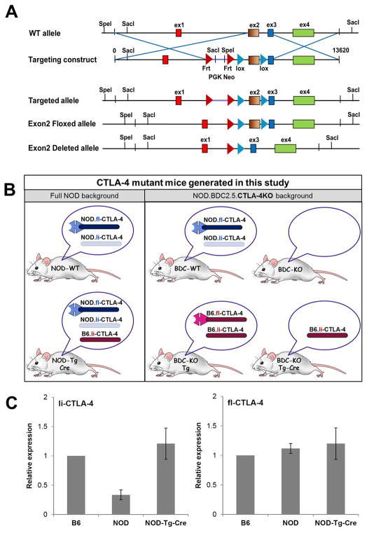
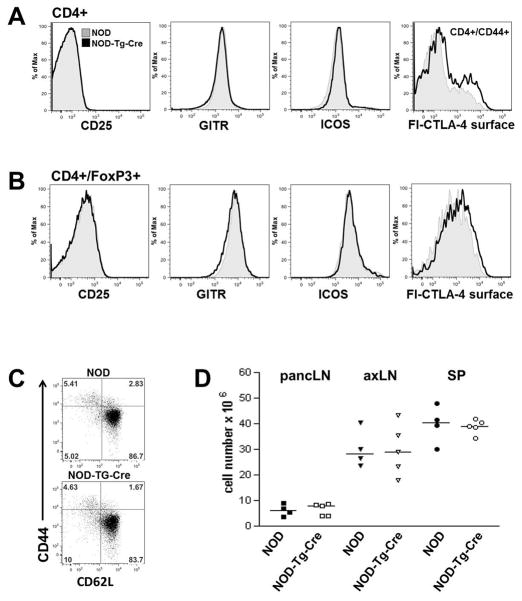
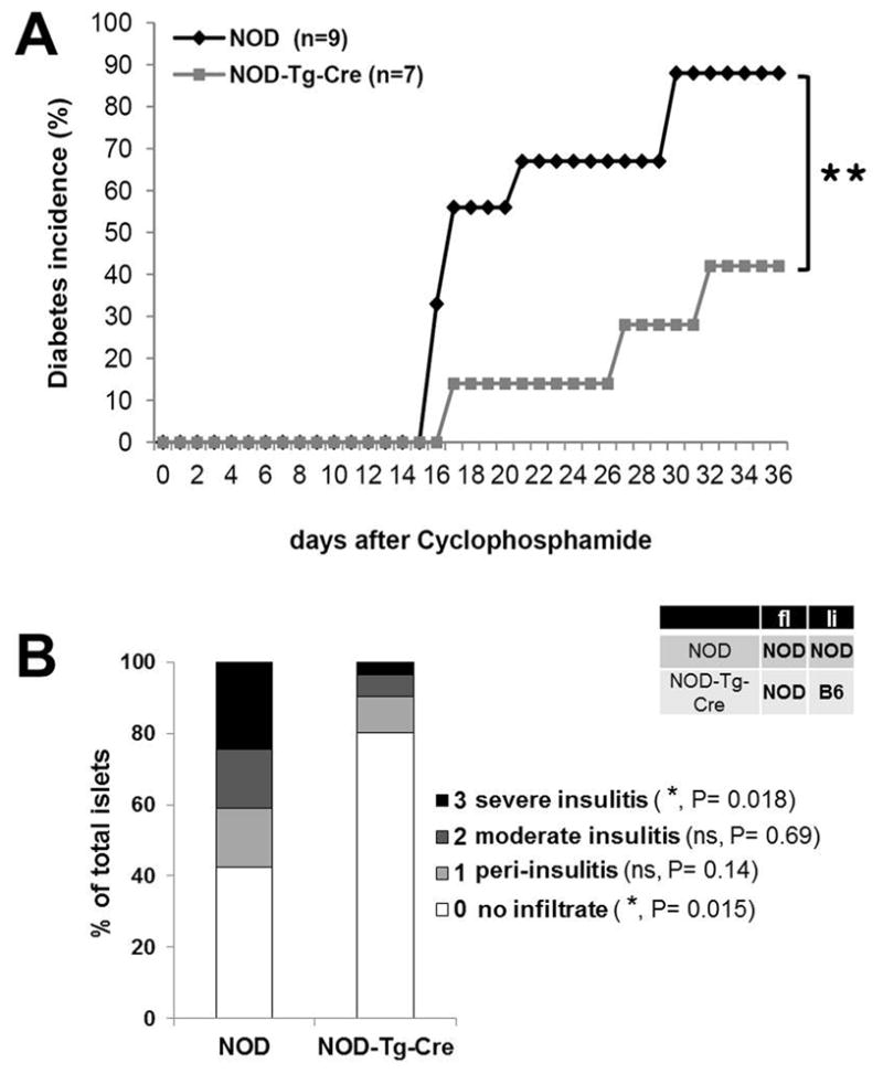
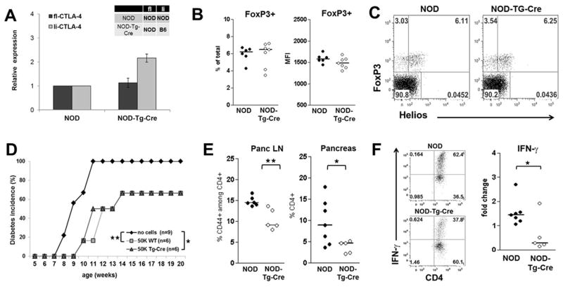
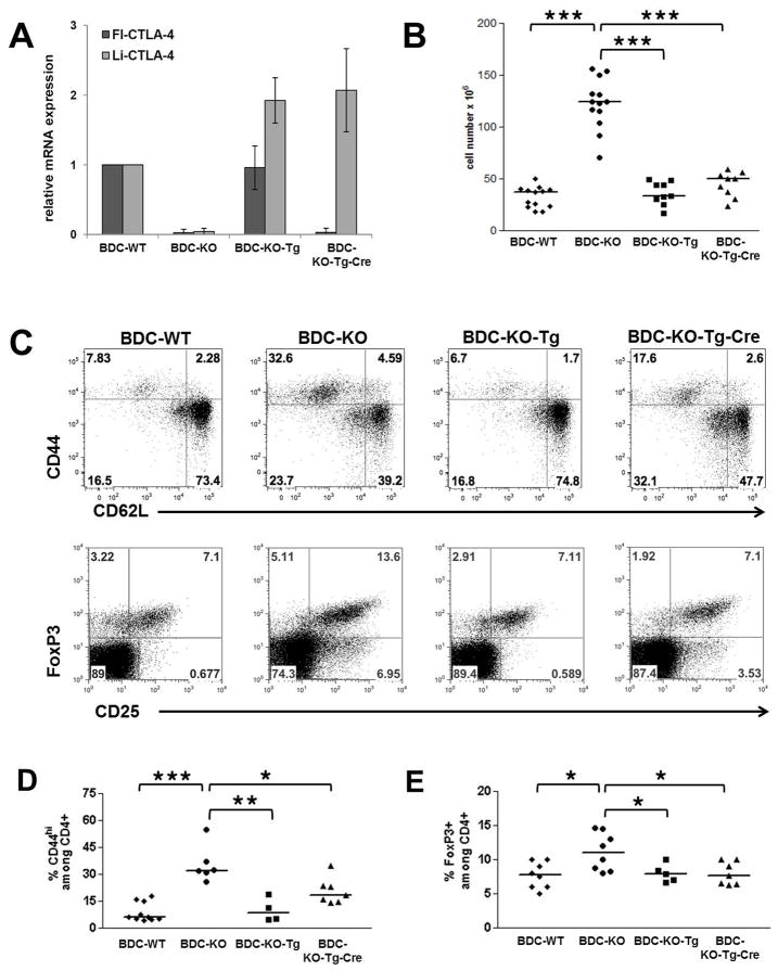
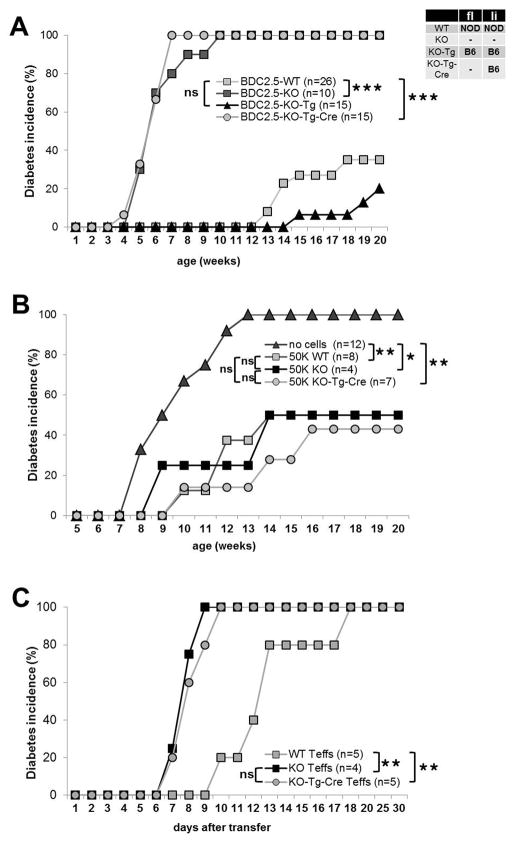
The critical role of CTLA-4 in inhibiting Ag-driven T cell responses upon engagement with its ligands, B7-1 and B7-2 and its importance for peripheral T cell tolerance and T cell homeostasis has been studied intensively. The CTLA-4 splice variant ligand-independent (li)-CTLA-4 is expressed in naive and activated T cells and can actively alter T cell signaling despite its lack of a B7 binding domain. To study the effect of li-CTLA-4 in regulating T cell responses in the context of autoimmunity, we engineered a B6.CTLA-4 (floxed-Exon2)-BAC-transgene, resulting in selective expression of li-CTLA-4 upon Cre-mediated deletion of Exon 2. Introducing the B6.BAC into the NOD background, which is genetically deficient for li-CTLA-4, restores mRNA levels of li-CTLA-4 to those observed in C57BL/6 mice. Furthermore, re-expressing this ligand nonbinding isoform in NOD mice reduced IFN-γ production in T effector cells accompanied by a significant decrease in insulitis and type 1 diabetes frequency. However, selective expression of li-CTLA-4 could not fully rescue the CTLA-4 knockout disease phenotype when bred onto NOD.BDC2.5.CTLA-4 knockout background because of the requirement of the full-length, B7-binding CTLA-4 molecule on T effector cells. Thus, the li-CTLA-4 form, when expressed at physiologic levels in the CTLA-4-sufficient NOD background can suppress autoimmunity; however, the functionality of the li-CTLA-4 isoform depends on the presence of the full-length molecule to alter effector T cell signaling.


Figures

Figure 1
Figure 1. Generation of CTLA-4 BAC-transgenic mice and expression of ligand-independent and full-length CTLA-4 isoforms
(A) Targeting strategy to generate CTLA-4 BAC-transgenic mice. A B6.BAC (RP24-316C5) containing exclusively the CTLA-4 locus was engineered to express 2 loxP sites, flanking Exon2 of the CTLA-4 gene. The modified B6.BAC was introduced into the NOD background and founders were crossed to the VAV-Cre deleter-strain to achieve excision of floxed Exon2, resulting in selective expression of only the B7, ligand non-binding isoform of CTLA-4. (B) Schematic representation of CTLA-4 mutant mice generated by introducing the B6.CTLA-4.BAC in the full NOD and NOD.BDC2.5 CTLA-4KO background. Expression of the full-length and or ligand-independent CTLA-4 isoforms from either the endogenous NOD or the B6-transgene allele is indicated for each mouse. (C) mRNA expression of full-length and ligand-independent CTLA-4 in CD4+ T cells of B6, NOD and NOD-Tg-Cre mice. Expression levels were normalized to 18S and are displayed as relative expression (mean ± SD).
Figure 2
Figure 2. T cell phenotype in prediabetic NOD mice expressing ligand-independent CTLA-4 at B6 levels
PancLN cells from 7 week old prediabetic wild-type NOD and NOD-Tg-Cre mice were stained for CD4, CD44, CD62L, CD25, GITR, ICOS and full-length CTLA-4 (fl-CTLA-4) to assess the activation status of CD4+ conventional (A), CD4+FoxP3+ regulatory T cells (B) and T effector/memory populations (C). (D) Total cellularity of pancreatic lymph node (pancLN), axillary lymph node (axLN) and spleen (SP) comparing 7 week old prediabetic wild-type NOD and NOD-Tg-Cre mice.
Figure 3
Figure 3. Reduced diabetes frequency in NOD mice expressing li-CTLA-4 at B6 levels
(A) Diabetes incidence in 8 week old NOD and NOD-Tg-Cre mice, injected intra peritoneal with 200mg/kg cyclophosphamide at days 0 and 7. Statistical analysis was performed using the Logrank test (**, P < 0.01). (B) Severity of pancreatic islet infiltration in 20 week old pre-diabetic NOD and NOD-Tg-Cre mice. Insulitis scores are representative for a total of 550 islets per group. Statistical analysis was performed using an unpaired t test.
Figure 4
Figure 4. Expression of li-CTLA-4 in NOD mice diminishes T effector cell function but does not affect T regulatory cells
(A) Relative levels of fl- and li-CTLA-4 mRNA expression normalized to 18S expression in sorted CD4+CD25hiCD62L Treg cells from NOD and NOD-Tg-Cre mice (mean ± SD). (B) Regulatory T cells numbers displayed as percentage of total pancLN cells (left panel) and FoxP3 protein expression levels (right panel). (C) Percentages of natural (CD4+FoxP3+Helios+) and adoptive (CD4+FoxP3+Helioslow) T regulatory cells in pancLN cells from NOD and NOD-Tg-Cre mice. (D) Diabetes frequency in NOD.CD28KO recipients adoptively transferred with either no or of 5×10⁴ BDC2.5-Treg cells from NOD and NOD-Tg-Cre mice, respectively. Statistical analysis was performed using the Logrank test (*, P < 0.05; **, P < 0.01). (E) Percentage of CD4+/CD44+ activated T effector cells in pancLN and pancreas of NOD and NOD-Tg-Cre mice. (F) Sorted CD4+/CD62Lhi T naïve cells from lymph nodes of NOD and NOD-Tg-Cre mice were activated with anti-CD3 and anti-CD28 in the presence of 200U/ml IL2. At day 3, cells were stimulated with PMA/Ionomycin/Monensin for 5 hours and the percentage of IFN-γ producing CD4+ T cells was examined by flow cytometry. Shown are representative flow plots (F, left panel) and the fold changes (F, right panel) of IFN-γ producing CD4+ T cells from NOD and NOD-Tg-Cre mice. Statistical analyses in E and F were performed using an unpaired t test (*, P < 0.05; **, P < 0.01).
Figure 5
Figure 5. Selective expression of li-CTLA-4 partially rescues the T cell phenotype observed in NOD.BDC2.5.CTLA-4KO mice
Relative levels of fl- and li-CTLA-4 mRNA expression in CD4+ T cells (A) and lymph node cellularity (B) in 6 week old NOD.BDC2.5.CTLA-4 WT, KO, KO-Tg or KO-Tg-Cre mice. (C) Representative flow plots, showing the frequencies of activated CD4+/CD44+/CD62L- T effector and resting CD4+/CD44-/CD62Lhi naïve T cells (upper panel) as well as CD4+/CD25+/FoxP3+ regulatory T cells (lower panel) in lymph node cells from 6 week old mice. (D) Quantification of frequencies of CD44+ T effector and FoxP3+ regulatory T cells determined in (C) and expressed as percentage of CD4+ T cells. Statistical analyses were performed using an unpaired t test (*, P < 0.05; **, P < 0.01; ***, P < 0.0001).
Figure 6
Figure 6. Selective expression of li-CTLA-4 in the NOD.BDC2.5.CTLA-4KO background does not protect from T1D development
(A) Diabetes incidence comparing NOD.BDC2.5.CTLA-4 WT, KO, KO-Tg or KO-Tg-Cre mice. (B) Diabetes frequency in NOD.CD28KO recipients adoptively transferred with either no or of 5×10⁴ BDC2.5-Treg cells from CTLA-4 WT, KO, KO-Tg or KO-Tg-Cre mice. (C) Diabetes development in full NOD recipients after adoptive transfer of 1×10⁶ in vitro activated BDC2.5-T effector cells from CTLA-4 WT, KO or KO-Tg-Cre mice, respectively. Statistical analyses were performed using the Logrank test (*, P < 0.05; **, P < 0.01; ***, P < 0.0001).

References

    1. Bour-Jordan H, Esensten JH, Martinez-Llordella M, Penaranda C, Stumpf M, Bluestone JA. Intrinsic and extrinsic control of peripheral T-cell tolerance by costimulatory molecules of the CD28/ B7 family. Immunol Rev. 2011;241:180–205. - PMC - PubMed
    1. Fife BT, Bluestone JA. Control of peripheral T-cell tolerance and autoimmunity via the CTLA-4 and PD-1 pathways. Immunol Rev. 2008;224:166–182. - PubMed
    1. Scalapino KJ, Daikh DI. CTLA-4: a key regulatory point in the control of autoimmune disease. Immunol Rev. 2008;223:143–155. - PubMed
    1. Gough SC, Walker LS, Sansom DM. CTLA4 gene polymorphism and autoimmunity. Immunol Rev. 2005;204:102–115. - PubMed
    1. Holmberg D, Cilio CM, Lundholm M, Motta V. CTLA-4 (CD152) and its involvement in autoimmune disease. Autoimmunity. 2005;38:225–233. - PubMed

Publication types

MeSH terms