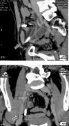
Barium appendicitis 1 month after a barium meal
- PMID: 23294068
- PMCID: PMC3727263
- DOI: 10.9738/CC160.1
Barium appendicitis 1 month after a barium meal
Abstract
Because barium sulfate (BaSO(4)) is not harmful to the mucosa, it is widely used for gastrointestinal imaging. Barium appendicitis is a very rare complication of barium meals and barium enema. We report a case of acute appendicitis associated with retained appendiceal barium. A 47-year-old man presented with right lower abdominal pain after upper gastrointestinal imaging was performed using barium 1 month earlier. The abdominal plain roentgenogram showed an area of retained barium in the right lower quadrant. Multiplanar reconstruction of computed tomography scans showed barium retention in the appendix. Emergency appendectomy was performed. A cross section of the specimen revealed the barium mass. Barium-associated appendicitis is a very rare clinical entity but we should be cautious of this uncommon disease when we encounter barium deposits in the appendix after barium examination. This report is significant because barium was identified both macroscopically and microscopically.
Figures


References
-
- Gubler JA, Kukral AJ. Barium appendicitis. J Int Col Surg. 1954;21(3:1):379–384. - PubMed
-
- Sisley JF, Wagner CW. Barium appendicitis. South Med J. 1982;75(4):498–499. - PubMed
-
- Fang YJ, Wang HP, Ho CM, Liu KL. Barium appendicitis. Surgery. 2009;21(9):957–958. - PubMed
-
- Novotny NM, Lillemore KD, Falimirski ME. Barium appendicitis after upper gastrointestinal imaging. J Emerg Med. 2010;38(2):148–149. - PubMed
-
- Cohen N, Modai D, Rosen A, Golic A, Weissgarten J. Barium appendicitis: fact or fancy? Report of a case and review of the literature. J Clin Gastroenterol. 1987;9(4):447–451. - PubMed
Publication types
MeSH terms
Substances
LinkOut - more resources
Full Text Sources
Other Literature Sources
Medical

