. 2013 Feb 6;135(5):1919-25.
doi: 10.1021/ja310901f. Epub 2013 Jan 25.

Optimized phospholipid bilayer nanodiscs facilitate high-resolution structure determination of membrane proteins

Affiliations

Optimized phospholipid bilayer nanodiscs facilitate high-resolution structure determination of membrane proteins

Franz Hagn et al. J Am Chem Soc. .

Abstract

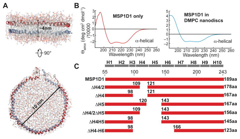
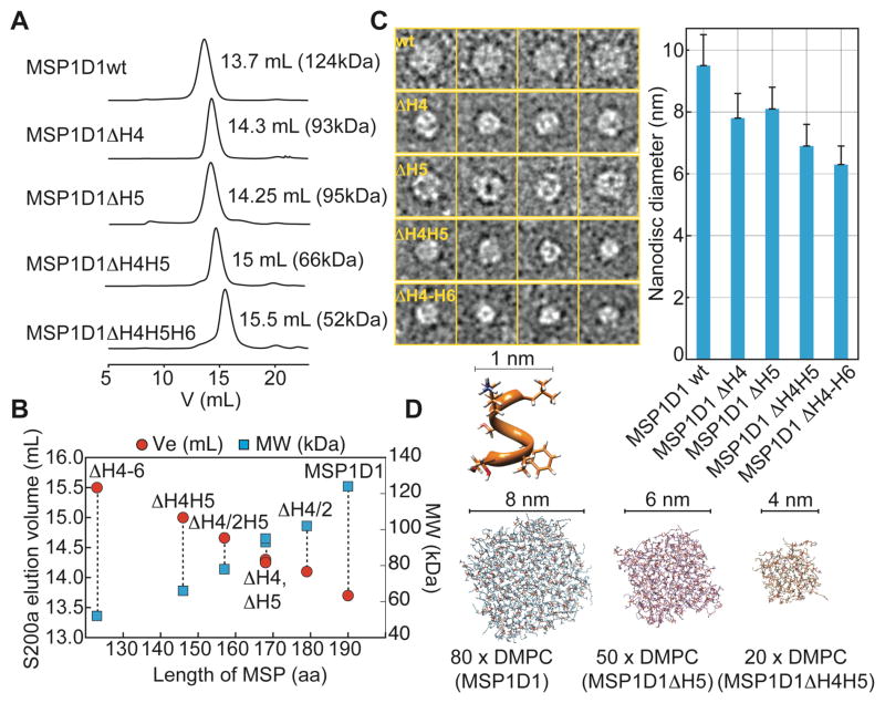
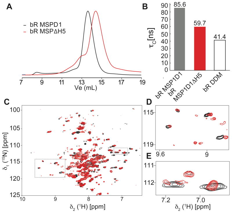
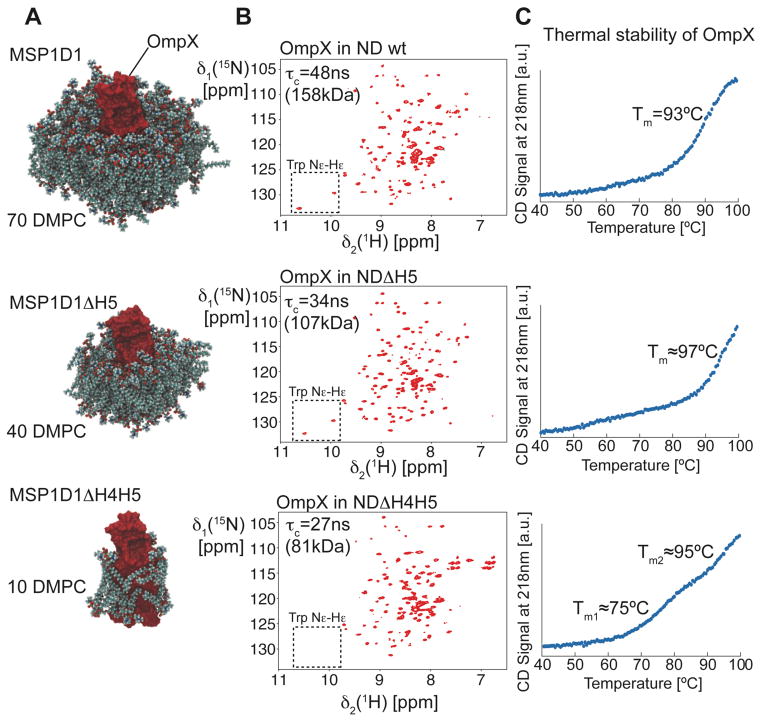
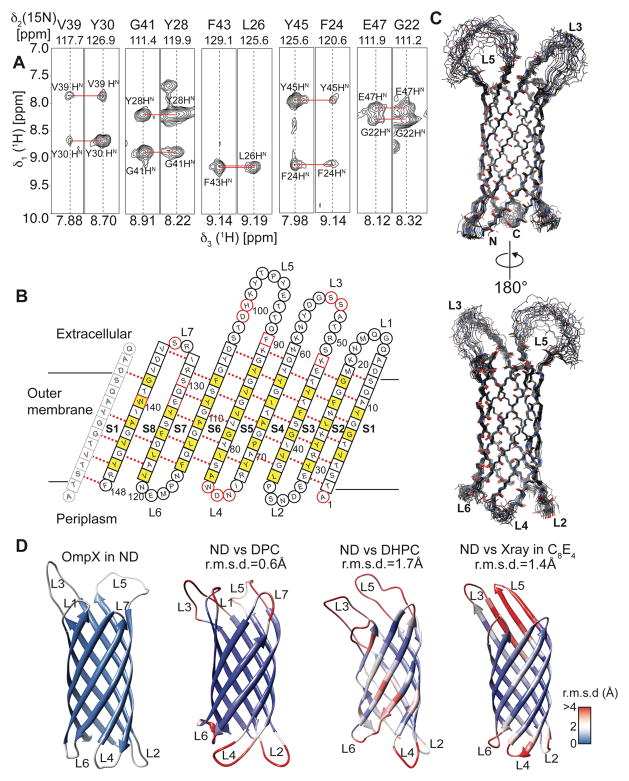
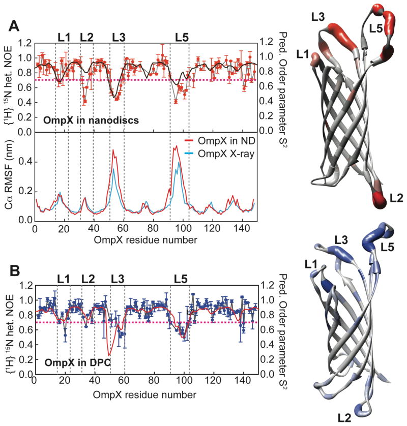
Structural studies of membrane proteins are still hampered by difficulties of finding appropriate membrane-mimicking media that maintain protein structure and function. Phospholipid nanodiscs seem promising to overcome the intrinsic problems of detergent-containing environments. While nanodiscs can offer a near-native environment, the large particle size complicates their routine use in the structural analysis of membrane proteins by solution NMR. Here, we introduce nanodiscs assembled from shorter ApoA-I protein variants that are of markedly smaller diameter and show that the resulting discs provide critical improvements for the structure determination of membrane proteins by NMR. Using the bacterial outer-membrane protein OmpX as an example, we demonstrate that the combination of small nanodisc size, high deuteration levels of protein and lipids, and the use of advanced non-uniform NMR sampling methods enable the NMR resonance assignment as well as the high-resolution structure determination of polytopic membrane proteins in a detergent-free, near-native lipid bilayer setting. By applying this method to bacteriorhodopsin, we show that our smaller nanodiscs can also be beneficial for the structural characterization of the important class of seven-transmembrane helical proteins. Our set of engineered nanodiscs of subsequently smaller diameters can be used to screen for optimal NMR spectral quality for small to medium-sized membrane proteins while still providing a functional environment. In addition to their key improvements for de novo structure determination, due to their smaller size these nanodiscs enable the investigation of interactions between membrane proteins and their (soluble) partner proteins, unbiased by the presence of detergents that might disrupt biologically relevant interactions.


Figures

Figure 1
Figure 1
Construction of truncated membrane scaffold protein (MSP) variants. (A) Proposed architecture of a phospholipid nanodisc where two copies of membrane scaffold protein (MSP) wrap around a patch of phospholipid bilayer, thereby stabilizing its hydrophobic edge. The most commonly used nanodisc has a diameter of 10 nm. Coordinates of the MSP were taken from ref.. (B) Far-UV CD spectra of MSP alone (left) or in an assembled nanodisc (right) show that MSP adopts α-helical secondary structure in both cases. (C) Deletion constructs of MSP1D1 used in this study. The predicted secondary structure of MSP1D1 is shown on top and the length of each construct is indicated.
Figure 2
Figure 2
Characterization of the smaller nanodiscs. (A) Size exclusion chromatograms of DMPC nanodiscs formed with various MSP1 deletion variants as indicated. The molecular weight was obtained by calibration of an analytical S200 column. (B) The elution volumes and the calculated molecular weights are linearly dependent on the length of the MSP protein that is in contact with lipids. (C) Representative negative-stain electron micrograms showing single nanodisc particles for the MSP variants as indicated. Right: Average diameter of nanodisc preparations determined by single particle analysis showing a decrease in nanodisc diameter from 9.5 to 6.3 nm with shorter MSP variants. Error bars indicate standard deviations (see also SI Fig. 2). (D) Assembly and equilibration of the experimentally determined number of DMPC lipids for MSP1D1, MSP1D1ΔH5 and MSP1D1ΔH4H5 by molecular dynamics simulations provide a fairly good estimate of the diameter of each nanodisc. To obtain the final diameter, one layer of the helical MSP protein (1 nm) was added to each side of the lipid bilayer.
Figure 3
Figure 3
Analysis of bacteriorhodopsin (bR) in different nanodiscs. (A) Analytical SEC traces of bR assembled into nanodiscs of different sizes. (B) Apparent rotational correlation time (τc) of bR in DMPC MSP1D1 (black) and MSP1D1ΔH5 (red) nanodiscs and in DDM micelles (white). (C) Overlay of 15N-1H-TROSY spectra of bR in MSP1D1 (black) and MSP1D1ΔH5 (red). (D,E) Expansion of boxed regions in (C).
Figure 4
Figure 4
Optimization of nanodisc size for the target protein OmpX. (A) Equilibrated box with OmpX and the number of DMPC lipids per bilayer leaflet determined experimentally. For clarity, the surrounding MSP protein is not shown. (B) 2D-[15N,1H]-TROSY experiments with 2H,15N-labeled OmpX in nanodiscs of different sizes, as indicated in the figure. The spectral region of the tryptophan side chain resonances is marked with a box. (C) Circular dichroism (CD)-detected thermal unfolding traces at 218nm of OmpX in different nanodiscs.
Figure 5
Figure 5
NMR backbone assignment and secondary structure determination of OmpX in MSP1D1ΔH5 nanodiscs. (A) 2D-[15N,1H]-TROSY spectrum of OmpX in MSP1D1ΔH5 DMPC/DPMG (3:1) nanodiscs at 45 °C and pH6.8. All assigned non-proline backbone amide resonances (90%) are labeled in the spectrum. (B) Example strips taken from the 3D-NUS HNCA and HN(CA)CB experiments show high spectral quality and resolution required for backbone resonance assignment. (C) Secondary structure prediction taken from the program Talos+ indicates 8 β-strands and long loop regions between strands 3 and 4 and strands 5 and 6.
Figure 6
Figure 6
Structure determination of OmpX in nanodiscs. (A) Example strips taken from a 3D-15N-edited-[1H,1H]-NOESY experiment showing long-range inter-β-strand contacts used for structure calculation. (B) Topology plot of OmpX. Residues facing towards the lipid bilayer are shown in yellow, unassigned residues in red. Residues in b-sheets are represented by squares whereas residues in loop regions are represented by circles. (C) The best-energy structural ensemble of (20 structures) of OmpX in nanodiscs has an r.m.s.d. of 0.32 Å between residues in β-sheets. (D) Comparison of the OmpX structure in nanodiscs (ND) with the NMR structures of OmpX in the detergents do-decylphosphocholine (DPC) (determined here), di-hexanoyl-glycero-phosphocholine (DHPC) and the crystal structure in n-octyltetraoxyethylene (C8E4). These structures are color-coded according to the r.m.s.d. values to individual residues of the nanodisc structure, as shown by the legend on the lower right. The r.m.s.d to ordered residues of the NMR structure in nanodiscs is indicated above each structure.
Figure 7
Figure 7
Dynamics of OmpX in nanodiscs and in DPC. (A) Upper left: {1H}-15N-heteronuclear NOE for OmpX in nanodiscs (red symbols) overlaid by the predicted squared order parameter S2 (black line). Right: Heteronuclear NOE values mapped onto the structure of OmpX (from grey to red). Lower left: Molecular dynamics simulations of OmpX in a phospholipid bilayer using the NMR structure obtained here (red line) or the previous X-ray structure (blue line) as starting conformations. The root mean square fluctuations (RMSF) of amino acid residues in OmpX are shown. Both simulations were run for 25 ns duration at 45ºC. Of note, even the well-ordered X-ray structure exhibits large fluctuations in the external long loop regions. (B) {1H}-15N-heteronuclear NOE for OmpX in DPC (blue symbols) overlaid by the predicted squared order parameter S2 (red line) based on chemical shift data. Right: Heteronuclear NOE values mapped onto the structure of OmpX in DPC (from grey to blue). Except at loop 2, these values are almost identical to the hetNOE obtained in nanodiscs. The dotted line at a hetNOE value of 0.7 represents the cutoff for coloring of residues in the structure.

References

    1. Linke D. Methods Enzymol. 2009;463:603. - PubMed
    1. Lee AG. Biochim Biophys Acta. 2003;1612:1. - PubMed
    1. Seddon AM, Curnow P, Booth PJ. Biochim Biophys Acta. 2004;1666:105. - PubMed
    1. Frauenfeld J, Gumbart J, Sluis EO, Funes S, Gartmann M, Beatrix B, Mielke T, Berninghausen O, Becker T, Schulten K, Beckmann R. Nat Struct Mol Biol. 2011;18:614. - PMC - PubMed
    1. Denisov IG, Grinkova YV, Lazarides AA, Sligar SG. J Am Chem Soc. 2004;126:3477. - PubMed

Publication types

MeSH terms