

High-frequency oscillations, extent of surgical resection, and surgical outcome in drug-resistant focal epilepsy
- PMID: 23294353
- PMCID: PMC3712982
- DOI: 10.1111/epi.12075
High-frequency oscillations, extent of surgical resection, and surgical outcome in drug-resistant focal epilepsy
Abstract
Purpose: Removal of areas generating high-frequency oscillations (HFOs) recorded from the intracerebral electroencephalography (iEEG) of patients with medically intractable epilepsy has been found to be correlated with improved surgical outcome. However, whether differences exist according to the type of epilepsy is largely unknown. We performed a comparative assessment of the impact of removing HFO-generating tissue on surgical outcome between temporal lobe epilepsy (TLE) and extratemporal lobe epilepsy (ETLE). We also assessed the relationship between the extent of surgical resection and surgical outcome.
Methods: We studied 30 patients with drug-resistant focal epilepsy, 21 with TLE and 9 with ETLE. Two thirds of the patients were included in a previous report and for these, clinical and imaging data were updated and follow-up was extended. All patients underwent iEEG investigations (500 Hz high-pass filter and 2,000 Hz sampling rate), surgical resection, and postoperative magnetic resonance imaging (MRI). HFOs (ripples, 80-250 Hz; fast ripples, >250 Hz) were identified visually on a 5-10 min interictal iEEG sample. HFO rates inside versus outside the seizure-onset zone (SOZ), in resected versus nonresected tissue, and their association with surgical outcome (ILAE classification) were assessed in the entire cohort, and in the TLE and ETLE subgroups. We also tested the correlation of resected brain hippocampal and amygdala volumes (as measured on postoperative MRIs) with surgical outcome.
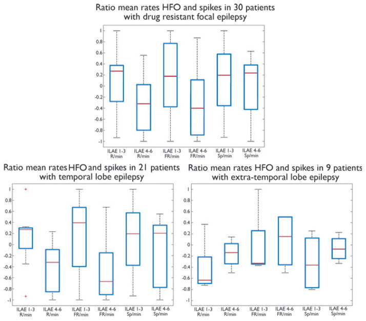
Key findings: HFO rates were significantly higher inside the SOZ than outside in the entire cohort and TLE subgroup, but not in the ETLE subgroup. In all groups, HFO rates did not differ significantly between resected and nonresected tissue. Surgical outcome was better when higher HFO rates were included in the surgical resection in the entire cohort and TLE subgroup, but not in the ETLE subgroup. Resected brain hippocampal and amygdala volumes were not correlated with surgical outcome.
Significance: In TLE, removal of HFO-generating areas may lead to improved surgical outcome. Less consistent findings emerge from ETLE, but these may be related to sample size limitations of this study. Size of resection, a factor that was ignored and that could have affected results of earlier studies did not influence results.
Wiley Periodicals, Inc. © 2012 International League Against Epilepsy.
Conflict of interest statement
None of the other authors has any conflict of interest to disclose. We confirm that we have read the Journal’s position on issues involved in ethical publication and affirm that this report is consistent with those guidelines.
Figures
References
-
- Akiyama T, McCoy B, Go CY, Ochi A, Elliott IM, Akiyama M, Donner EJ, Weiss SK, Snead OC, III, Rutka JT, Drake JM, Otsubo H. Focal resection of fast ripples on extraoperative intracranial EEG improves seizure outcome in pediatric epilepsy. Epilepsia. 2011;52:1802–1811. - PubMed
-
- Bénar CG, Chauvière L, Bartolomei F, Wendling F. Pitfalls of high-pass filtering for detecting epileptic oscillations: a technical note on “false” ripples. Clin Neurophysiol. 2010;121:301–310. - PubMed
-
- Bernasconi A, Bernasconi N. Unveiling epileptogenic lesions: the contribution of image processing. Epilepsia. 2011;52(Suppl 4):20–24. - PubMed
-
- Bragin A, Engel J, Jr, Wilson CL, Fried I, Mathern GW. Hippocampal and entorhinal cortex high-frequency oscillations (100–500 Hz) in human epileptic brain and in kainic acid-treated rats with chronic seizures. Epilepsia. 1999a;40:127–137. - PubMed
Publication types
MeSH terms
Grants and funding
LinkOut - more resources
Full Text Sources
Other Literature Sources
