. 2013 Mar 19;46(3):681-91.
doi: 10.1021/ar3000647. Epub 2013 Jan 7.

The role of surface functionality in determining nanoparticle cytotoxicity

Affiliations

The role of surface functionality in determining nanoparticle cytotoxicity

Sung Tae Kim et al. Acc Chem Res. .

Abstract

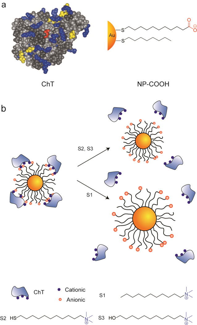
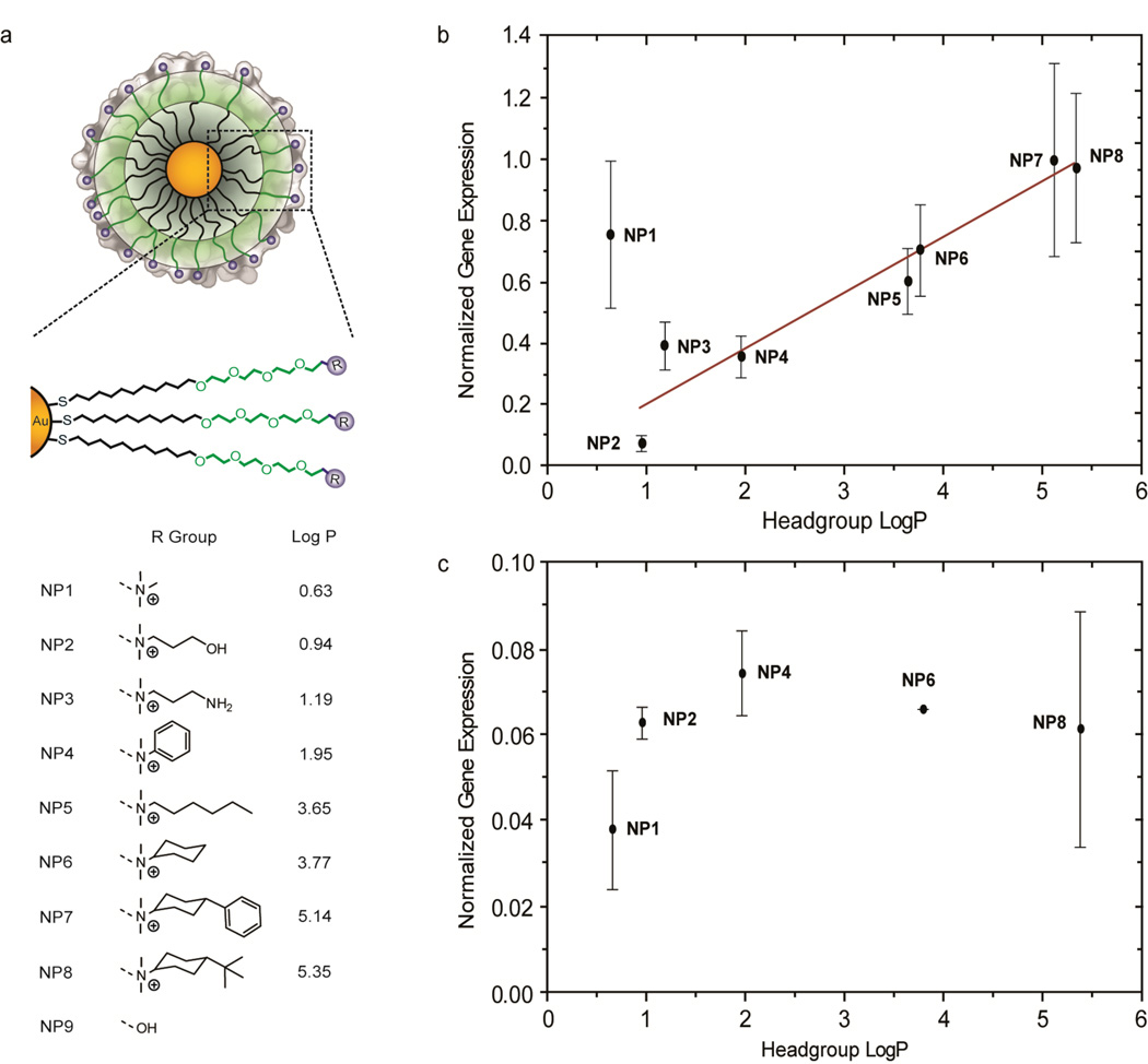
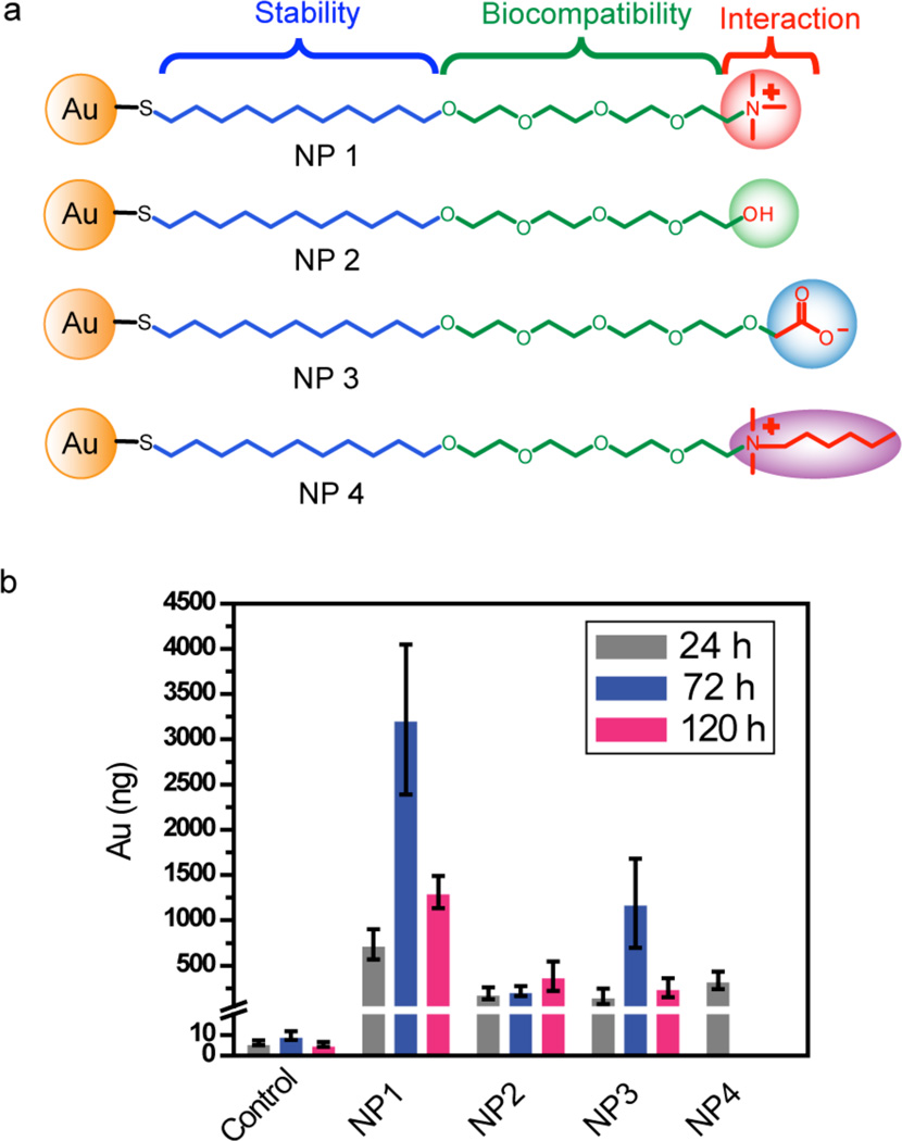
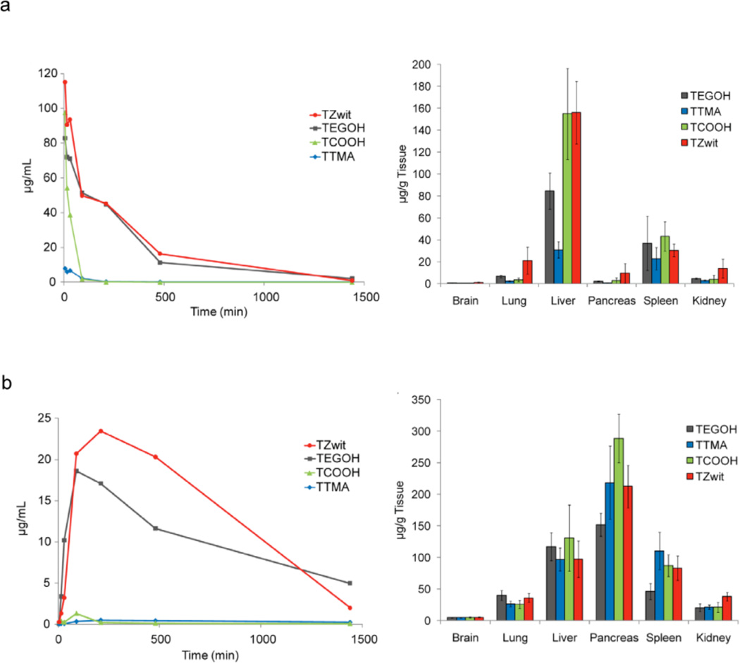
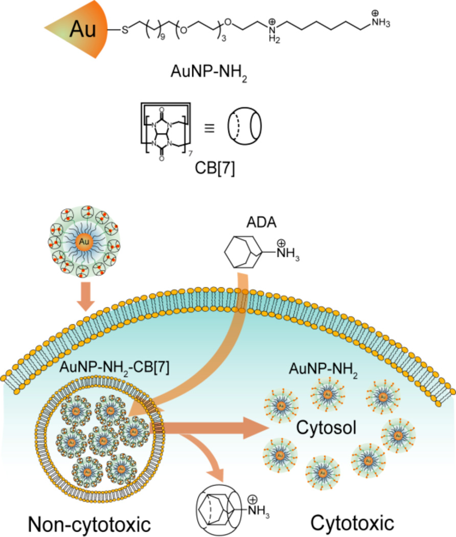
Surface properties dictate the behavior of nanomaterials in vitro, in vivo, and in the environment. Such properties include surface charge and hydrophobicity. Also key are more complex supramolecular interactions such as aromatic stacking and hydrogen bonding, and even surface topology from the structural to the atomic level. Surface functionalization of nanoparticles (NPs) provides an effective way to control the interface between nanomaterials and the biological systems they are designed to interact with. In medicine, for instance, proper control of surface properties can maximize therapeutic or imaging efficacy while minimizing unfavorable side effects. Meanwhile, in environmental science, thoughtful choice of particle coating can minimize the impact of manufactured nanomaterials on the environment. A thorough knowledge of how NP surfaces with various properties affect biological systems is essential for creating NPs with such useful therapeutic and imaging properties as low toxicity, stability, biocompatibility, favorable distribution throughout cells or tissues, and favorable pharmacokinetic profiles--and for reducing the potential environmental impact of manufactured nanomaterials, which are becoming increasingly prominent in the marketplace. In this Account, we discuss our research and that of others into how NP surface properties control interactions with biomolecules and cells at many scales, including the role the particle surface plays in determining in vivo behavior of nanomaterials. These interactions can be benign, beneficial, or lead to dysfunction in proteins, genes and cells, resulting in cytotoxic and genotoxic responses. Understanding these interactions and their consequences helps us to design minimally invasive imaging and delivery agents. We also highlight in this Account how we have fabricated nanoparticles to act as therapeutic agents via tailored interactions with biomacromolecules. These particles offer new therapeutic directions from traditional small molecule therapies, and with potentially greater versatility than is possible with proteins and nucleic acids.


Figures

Figure 1
Figure 1
Effect of functionalized NPs on the disruption of lipid bilayers. (a) Surface functionalized MMPC1 and MMPC2 and (b) Comparison of cationic MMPC1 and anionic MMPC2 (220 nM) in disrupting vesicles with an overall negative charge (SOPC/SOPC, L-R-stearoyloleoyl-phosphatidylcholine/L-R-stearoyl-oleoyl-phosphatidylserine). Reprinted with permission from ref.. Copyright 2004 American Chemical Society.
Figure 2
Figure 2
Effect of gold NPs with different surface charges on cellular membrane potential. (a) Cationic, anionic, neutral, and zwitterionic NPs. (b) Membrane potential changes following the exposure to NPs for ovarian cancer cells (CP70 and A2780), human bronchial epithelial cells (BEC), and human airway smooth muscle cells (ASM) using cell permeable fluorescent membrane potential indicator RH414 and real-time fluorescence microscopy. In addition, the extent of membrane potential change was analyzed in a cationic NP concentration dependent manner. (*p < 0.05) (c) Scheme of NP effects on cell and TEM of cationic NP interactions with plasma membrane. Reprinted with permission from ref.. Copyright 2010 American Chemical Society.
Figure 3
Figure 3
Interaction between positively charged NPs and DNA. (a) Mixed monolayer protected gold clusters (MMPCs) and double stranded DNA (37mer). (b) The amount of RNA detected relative to levels produced in the absence of MMPCs. Reprinted with permission from ref.. Copyright 2001, American Chemical Society.
Figure 4
Figure 4
Cytotoxicity and genotoxicity of gold NPs with different hydrophobicities. (a) Gold NPs (e.g. NP-TTMA, -ET, -BU and -HEX) (b) IC50 values of these NPs were determined by alamarBlue® assay. (c) ROS was quantitatively determined by the oxidation of 2′,7′-dichlorodihydrofluorescein diacetate dye. (d) Tail length and % Tail DNA were measured by the Comet assay. Reprinted with permission from ref.. Copyright 2010 Wiley-VCH Verlag & Co. KGaA.
Figure 5
Figure 5
Reversible/irreversible interaction of ChT with surface functionality of gold NP. (a) Space-filling model of ChT and structure of anionic MUA-gold NP (MUA-NP) (b) ChT released from the surface of NP by the addition of different trimethylamine-functionalized surfactants (S1, S2 and S3). Reprinted with permission from ref.. Copyright 2003, American Chemical Society.
Figure 6
Figure 6
Effect of NP surface hydrophobicity on gene expression related to immune response. (a) Surface functionalized gold NPs controlling the surface hydrophobicity and cytokine gene expression of (b) TNF- α in vitro and (c) IL-10 in vivo as function of NP headgroup LogP. LogP represents the calculated hydrophobic values of the head group. Reprinted with permission from ref.. Copyright 2012, American Chemical Society.
Figure 7
Figure 7
Effect of NPs with different functionalities on total accumulation of gold in Japanese medaka fish. (a) Gold NPs with different functionalities and (b) total amount of gold detected in fish after exposure of 20 nM gold NP concentrations. The gold amounts are the sum total of gold found in various organs (brain, heart, liver, gonads, gills, intestines and dorsal fin) and on appendage. Reprinted with permission from ref.. Copyrights 2010 Wiley-VCH Verlag & Co. KGaA.
Figure 8
Figure 8
Pharmacokinetic profiles (left panel) and biodistribution (right panel) of gold NPs in vivo. Four kinds of NPs (TEGOH, TTMA, TCOOH and TZwit) were administered into mice via (a) intravenous or (b) intraperitoneal route. Pharmacokinetic studies were performed for 1 day in normal male CD1 mice and organs were collected 1 day after administrations from ovarian cancer cell (CP-70) transplanted HEJ/C3H mice. Adapted from ref..
Figure 9
Figure 9
Schematic description of the use of intracellular host–guest complexation to trigger gold nanoparticle cytotoxicity. Cytotoxicity of gold NP-NH2-CB[7] is activated by the dethreading of CB[7] from the nanoparticle surface by ADA. Reprinted with permission from ref.. Copyright 2010 Nature Publishing Group.

References

    1. Smith AM, Duan HW, Mohs AM, Nie SM. Bioconjugated quantum dots for in vivo molecular and cellular imaging. Adv. Drug Delivery Rev. 2008;60:1226–1240. - PMC - PubMed
    1. De M, Rana S, Akpinar H, Miranda OR, Arvizo RR, Bunz UHF, Rotello VM. Sensing of proteins in human serum using conjugates of nanoparticles and green fluorescent protein. Nat. Chem. 2009;1:461–465. - PMC - PubMed
    1. Davis ME, Chen Z, Shin DM. Nanoparticle therapeutics: an emerging treatment modality for cancer. Nat. Rev. Drug Discov. 2008;7:771–782. - PubMed
    1. Gates BC. Catalysis: Individual nanoparticles in action. Nat. Nanotechnol. 2008;3:583–584. - PubMed
    1. Weickert J, Dunbar RB, Hesse HC, Wiedemann W, Schmidt-Mende L. Nanostructured organic and hybrid solar cells. Adv. Mater. 2011;23:1810–1828. - PubMed

Publication types