

Fluorescence resonance energy transfer glucose sensor from site-specific dual labeling of glucose/galactose binding protein using ligand protection
- PMID: 23294773
- PMCID: PMC3570868
- DOI: 10.1177/193229681200600607
Fluorescence resonance energy transfer glucose sensor from site-specific dual labeling of glucose/galactose binding protein using ligand protection
Abstract
Background: Site-selective modification of proteins at two separate locations using two different reagents is highly desirable for biosensor applications employing fluorescence resonance energy transfer (FRET), but few strategies are available for such modification. To address this challenge, sequential selective modification of two cysteines in glucose/galactose binding protein (GGBP) was demonstrated using a technique we call "ligand protection."
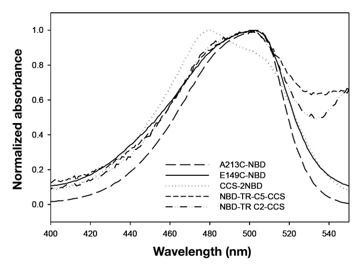
Method: In this technique, two cysteines were introduced in GGBP and one cysteine is rendered inaccessible by the presence of glucose, thus allowing sequential attachment of two different thiol-reactive reagents. The mutant E149C/A213C/L238S was first labeled at E149C in the presence of the ligand glucose. Following dialysis and removal of glucose, the protein was labeled with a second dye, either Texas Red (TR) C5 bromoacetamide or TR C2 maleimide, at the second site, A213C.
Results: Changes in glucose-dependent fluorescence were observed that were consistent with FRET between the nitrobenzoxadiazole and TR fluorophores. Comparison of models and spectroscopic properties of the C2 and C5 TR FRET constructs suggests the greater rigidity of the C2 linker provides more efficient FRET.
Conclusions: The ligand protection strategy provides a simple method for labeling GGBP with two different fluorophores to construct FRET-based glucose sensors with glucose affinity within the human physiological glucose range (1-30 mM). This general strategy may also have broad utility for other protein-labeling applications.
© 2012 Diabetes Technology Society.
Figures


References
-
- Geoghegan KF, Stroh JG. Site-directed conjugation of nonpeptide groups to peptides and proteins via periodate oxidation of a 2-amino alcohol. Application to modification at N-terminal serine. Bioconjug Chem. 1992;3(2):138–146. - PubMed
-
- Gaertner HF, Offord RE. Site-specific attachment of functionalized poly(ethylene glycol) to the amino terminus of proteins. Bioconjug Chem. 1996;7(1):38–44. - PubMed
-
- Zhao ZG, Im JS, Lam KS, Lake DF. Site-specific modification of a single-chain antibody using a novel glyoxylyl-based labeling reagent. Bioconjug Chem. 1999;10(3):424–430. - PubMed
-
- Wood RJ, Pascoe DD, Brown ZK, Medlicott EM, Kriek M, Neylon C, Roach PL. Optimized conjugation of a fluorescent label to proteins via intein-mediated activation and ligation. Bioconjug Chem. 2004;15(2):366–372. - PubMed
-
- Turcatti G, Nemeth K, Edgerton MD, Meseth U, Talabot F, Peitsch M, Knowles J, Vogel H, Chollet A. Probing the structure and function of the tachykinin neurokinin-2 receptor through biosynthetic incorporation of fluorescent amino acids at specific sites. J Biol Chem. 1996;271(33):19991–19998. - PubMed
Publication types
MeSH terms
Substances
LinkOut - more resources
Full Text Sources
Other Literature Sources
Miscellaneous
