

3D engineered cardiac tissue models of human heart disease: learning more from our mice
- PMID: 23295081
- PMCID: PMC4066654
- DOI: 10.1016/j.tcm.2012.08.007
3D engineered cardiac tissue models of human heart disease: learning more from our mice
Abstract
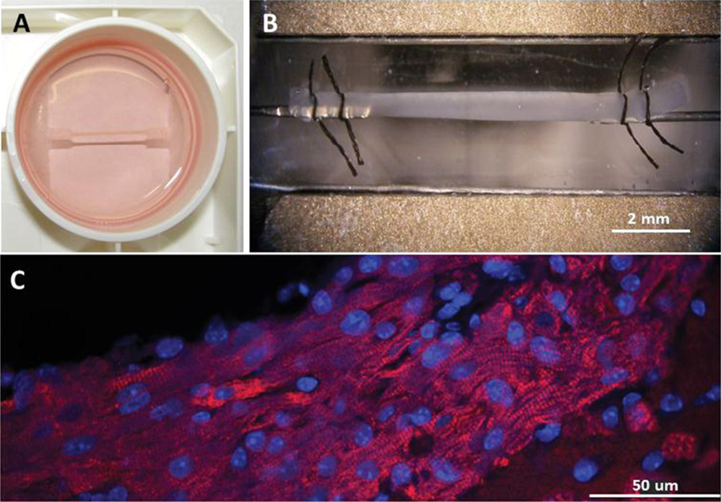
Mouse engineered cardiac tissue constructs (mECTs) are a new tool available to study human forms of genetic heart disease within the laboratory. The cultured strips of cardiac cells generate physiologic calcium transients and twitch force, and respond to electrical pacing and adrenergic stimulation. The mECT can be made using cells from existing mouse models of cardiac disease, providing a robust readout of contractile performance and allowing a rapid assessment of genotype-phenotype correlations and responses to therapies. mECT represents an efficient and economical extension to the existing tools for studying cardiac physiology. Human ECTs generated from iPSCMs represent the next logical step for this technology and offer significant promise of an integrated, fully human, cardiac tissue model.
Copyright © 2013. Published by Elsevier Inc.
Figures
References
-
- Bell RM, Mocanu MM, Yellon DM. Retrograde heart perfusion: the Langendorff technique of isolated heart perfusion. Journal of Molecular and Cellular Cardiology. 2011;50:940–950. - PubMed
-
- Chu G, Haghighi K, Kranias EG. From mouse to man: understanding heart failure through genetically altered mouse models. Journal of Cardiac Failure. 2002;8:S432–S449. - PubMed
-
- Eschenhagen T, Fink C, Remmers U, et al. Three-dimensional reconstitution of embryonic cardiomyocytes in a collagen matrix: a new heart muscle model system. FASEB Journal. 1997;11:683–694. - PubMed
Publication types
MeSH terms
Grants and funding
LinkOut - more resources
Full Text Sources
Other Literature Sources
Medical
