. 2012 Dec;67(12):1379-86.
doi: 10.6061/clinics/2012(12)06.

Mechanical thrombectomy with solitaire stent retrieval for acute ischemic stroke in a Brazilian population

Affiliations

Mechanical thrombectomy with solitaire stent retrieval for acute ischemic stroke in a Brazilian population

Luis Henrique de Castro-Afonso et al. Clinics (Sao Paulo). 2012 Dec.

Abstract

Objective: Large vessel occlusion in acute ischemic stroke is associated with low recanalization rates under intravenous thrombolysis. We evaluated the safety and efficacy of the Solitaire AB stent in treating acute ischemic stroke.

Methods: Patients presenting with acute ischemic stroke were prospectively evaluated. The neurological outcomes were assessed using the National Institutes of Health Stroke Scale and the modified Rankin Scale. Time was recorded from the symptom onset to the recanalization and procedure time. Recanalization was assessed using the thrombolysis in cerebral infarction score.

Results: Twenty-one patients were evaluated. The mean patient age was 65, and the National Institutes of Health Stroke Scale scores ranged from 7 to 28 (average 17 ± 6.36) at presentation. The vessel occlusions occurred in the middle cerebral artery (61.9%), distal internal carotid artery (14.3%), tandem carotid occlusion (14.3%), and basilarartery (9.5%). Primary thrombectomy, rescue treatment and a bridging approach represented 66.6%, 28.6%, and 4.8% of the performed procedures, respectively. The mean time from symptom onset to recanalization was 356.5 ± 107.8 minutes (range, 80-586 minutes). The mean procedure time was 60.4 ± 58.8 minutes (range, 14-240 minutes). The overall recanalization rate (thrombolysis in cerebral infarction scores of 3 or 2b) was 90.4%, and the symptomatic intracranial hemorrhage rate was 14.2%. The National Institutes of Health Stroke Scale scores at discharge ranged from 0 to 25 (average 6.9 ± 7). At three months, 61.9% of the patients had a modified Rankin Scale score of 0 to 2, with an overall mortality rate of 9.5%.

Conclusions: Intra-arterial thrombectomy with the Solitaire AB device appears to be safe and effective. Large randomized trials are necessary to confirm the benefits of this approach in acute ischemic stroke.


Conflict of interest statement

No potential conflict of interest was reported.

Figures

Figure 1
Figure 1
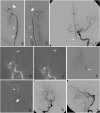
(a) Digital subtraction angiography (DSA) of the right common carotid artery ([RCCA] arterial phase, oblique view) shows an occlusion of the distal right internal carotid artery (RICA) (black arrow); (b) a road map of the RICA (oblique view) shows a guiding catheter in the RICA (arrowhead); the distal tip of the microcatheter (white arrow) and the microwire have crossed the occluded portion of the RICA and moved through the thrombi and into the right middle cerebral artery (RMCA) (black arrow); (c) a road map of the RICA (oblique view) shows the terminal tip of microcatheter (white arrow) distal to the occlusion site; (d) a road map of the RICA (oblique view) shows the terminal tip of the microcatheter (white arrow) and the terminal radiopaque marks of a 6x30 mm Solitaire AB device (ev3, Irvine, CA, USA), which indicate the start of deployment (white arrowhead); (e) a road map of the RICA (oblique view) in which the proximal stent radiopaque mark is seen ahead of the distal tip of microcatheter (white arrowhead), indicating the full deployment of the stent into the RMCA and the distal RICA; (f) a control DSA of the RCCA (arterial phase, oblique view) that was performed after the stent retrieval shows that the distal RICA, right anterior cerebral artery (RACA) and RMCA branches have been completely opened; (g) a picture shows the Solitaire AB (ev3, Irvine, CA, USA) and the removed clot.
Figure 2
Figure 2
(a) DSA of the left common carotid artery (LCCA), arterial phase, frontal view, shows a proximal occlusion of M1 segment of left middle cerebral artery (LMCA) (black arrow); (b) DSA of left interna carotid artery (LICA), arterial phase, frontal view, shows a Solitaire AB (ev3, Irvine, CA, USA) deployed into the M1 segment and promptly flow restoration of LMCA (black arrow); (c) Final control DSA of the LCCA, arterial phase, frontal view, shows the complete recanalization of the LMCA; (d, e, f) road maps of LICA, frontal views, show (d) the microwire crossed the occluded LMCA (black arrow), (e) the terminal tip of the microcatheter (black arrow) placed beyond the thrombi into the M1 segment of the LMCA and (f) terminal radiopaque marks of a 4x20 mm Solitaire AB device (ev3) deployed into the occluded site of LMCA; (g, h, i) Brain CT scans, (g) performed on presentation revealing no signs of hemorrhage or ischemic injury, (h) performed immediately after intra-arterial thrombectomy showing contrast medium enhance in the left middle cerebral artery territory and (i) performed 48 hours after intra-arterial thrombectomy showing contrast medium cleared and slight parenchymal hypoattenuation signs.
Figure 3
Figure 3
(a, b) a DSA of (a) the right vertebral artery (RVA) and (b) the left vertebral artery (LVA) (white arrows), arterial phase, frontal views, shows an occlusion of the proximal basilar artery (BA) (white arrowheads); (c) a DSA of the LCCA (frontal view) shows the BA (white arrow) filled by the left posterior communicating artery; (d, e) a Road map of the RVA (frontal view) shows (d) the microwire (white arrow) crossing the occluded section of the BA and (e) the distal tip of the microcatheter (white arrow) advanced by the microwire through the thrombi; (f) a DSA of the BA (frontal view) shows the contrast medium being injected through the microcatheter distal to the thrombi in the BA (white arrow); (g) the distal (white arrow) and proximal (white arrowhead) radiopaque marks of the stent (ev3, Irvine, CA, USA) reveal its full deployment into the distal RVA and BA; (h, i) A final control DSA of the RVA (arterial phase, [h] frontal and [i] lateral views) shows the complete recanalization of the BA.

References

    1. The World Health Report 2008. Geneva; World Health Organization. 2008
    1. Ministério da Saúde. Mortalidade–Brasil. Óbitos por Ocorrência por Sexo Segundo Causa CID–BR–10. Accessed May 01, 2010.
    1. Christensen MC, Valiente R, Sampaio Silva G, Lee WC, Dutcher S, Guimarães Rocha MS, et al. Acute treatment costs of stroke in Brazil. Neuroepidemiology. 2009;32(2):142–9. - PubMed
    1. Pontes-Neto OM, Silva GS, Feitosa MR, de Figueiredo NL, Fiorot JA, Jr, Rocha TN, et al. Stroke awareness in Brazil: alarming results in a community-based study. Stroke. 2008;39(2):292–6. - PubMed
    1. Massaro AR. Stroke in Brazil: a South America perspective. Int J Stroke. 2006;1(2):113–5. - PubMed