Review
. 2013 Jan 8;80(2):210-9.
doi: 10.1212/WNL.0b013e31827b910b.

The thalamus and multiple sclerosis: modern views on pathologic, imaging, and clinical aspects

Affiliations
Review

The thalamus and multiple sclerosis: modern views on pathologic, imaging, and clinical aspects

Alireza Minagar et al. Neurology. .

Abstract

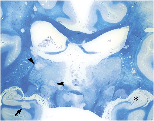
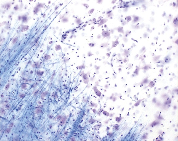
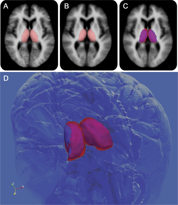
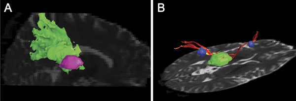
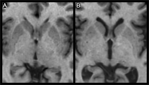
The paired thalamic nuclei are gray matter (GM) structures on both sides of the third ventricle that play major roles in cortical activation, relaying sensory information to the higher cortical centers that influence cognition. Multiple sclerosis (MS) is an immune-mediated disease of the human CNS that affects both the white matter (WM) and GM. A number of clinical observations as well as recent neuropathologic and neuroimaging studies have clearly demonstrated extensive involvement of the thalamus, basal ganglia, and neocortex in patients with MS. Modern MRI techniques permit visualization of GM lesions and measurement of atrophy. These contemporary methods have fundamentally altered our understanding of the pathophysiologic nature of MS. Evidence confirms the contention that GM injury can be detected in the earliest phases of MS, and that iron deposition and atrophy of deep gray nuclei are closely related to the magnitude of inflammation. Extensive involvement of GM, and particularly of the thalamus, is associated with a wide range of clinical manifestations including cognitive decline, motor deficits, fatigue, painful syndromes, and ocular motility disturbances in patients with MS. In this review, we characterize the neuropathologic, neuroimaging, and clinical features of thalamic involvement in MS. Further, we underscore the contention that neuropathologic and neuroimaging correlative investigations of thalamic derangements in MS may elucidate not heretofore considered pathobiological underpinnings germane to understanding the ontogeny, magnitude, and progression of the disease process.


Figures

Figure 1
Figure 1. Coronal section through the posterior thalamus, secondary progressive multiple sclerosis
Focal chronic lesions are present in the right pulvinar nuclei and the lateral posterior nucleus of the thalamus (arrowheads), the latter with a remyelinating fringe. Demyelination is also present in the deep gray matter of the right caudate nucleus in association with an adjacent chronic white matter lesion. Additionally, there is a leukocortical lesion in the subiculum of the right hippocampal formation (arrow), diffuse demyelination of the left hippocampus proper (asterisk), and involvement of the periaqueductal gray matter. Classic small focal white matter lesions are present in the white matter, including the corpus callosum. Luxol fast blue.
Figure 2
Figure 2. Border zone of a chronic thalamic lesion, secondary progressive multiple sclerosis
The gray matter lesion (top right) is devoid of myelin (stained blue) and hypocellular. Comparison with adjacent normal-appearing gray matter (bottom left) discloses a reduction in the number of neurons and a paucity of cells with oligodendrocyte morphology (Luxol fast blue).
Figure 3
Figure 3. Comparison of thalamic segmentations
Comparison of thalamic segmentations between 26 healthy controls (HC) (age- and sex-matched) (A) and 98 patients with relapsing-remitting multiple sclerosis (MS) (B). (C) Voxel-wise differences between the groups, with magenta representing common areas, red HC-only areas, and blue MS-only areas. Individual patient images were coregistered into the Montreal Neurological Institute 152 space for visualization. A 3D view (D) shows that the MS thalami are smaller overall and slightly shifted away from the midline (most likely by global atrophic processes).
Figure 4
Figure 4. Representative 3D rendering example of fiber tracking from diffusion tensor imaging and voxel-based morphometry in multiple sclerosis
(A) Example of a probabilistic thalamoparietal template. The Montreal Neurological Institute transformed map is coregistered into the patient space; the thalamoparietal region of interest (ROI) is in green and the thalamus ROI in pink. (B) The figure shows 2 thalamocortical fiber tracks (red) seeded through white matter lesions (blue) connecting the thalamus (green) and the cortex (not shown).
Figure 5
Figure 5. Thalamic atrophy and third ventricle width in multiple sclerosis
Thalamic atrophy is closely related to third ventricle width, as seen in these images of a patient with relapsing-remitting multiple sclerosis (MS) (A) and a patient with secondary progressive MS (B). Both patients have a disease duration of 14 years, and both images are of the same slice in stereotactic Montreal Neurological Institute 152 space.

References

    1. Kutzelnigg A, Lassmann H. Cortical lesions and brain atrophy in MS. J Neurol Sci 2005;233:55–59 - PubMed
    1. Wegner C, Esiri MM, Chance SA, Palace J, Matthews PM. Neocortical neuronal, synaptic, and glial loss in multiple sclerosis. Neurology 2006;67:960–967 - PubMed
    1. Lucchinetti CF, Popescu BF, Bunyan RF, et al. Inflammatory cortical demyelination in early multiple sclerosis. N Engl J Med 2011;365:2188–2197 - PMC - PubMed
    1. Dutta R, Chang A, Doud MK, et al. Demyelination causes synaptic alterations in hippocampi from multiple sclerosis patients. Ann Neurol 2011;69:445–454 - PMC - PubMed
    1. Popescu BF, Lucchinetti CF. Meningeal and cortical grey matter pathology in multiple sclerosis. BMC Neurol 2012;12:11. - PMC - PubMed