. 2013 Jan 22;110(4):1339-44.
doi: 10.1073/pnas.1221538110. Epub 2013 Jan 7.

Calcium oscillations-coupled conversion of actin travelling waves to standing oscillations

Affiliations

Calcium oscillations-coupled conversion of actin travelling waves to standing oscillations

Min Wu et al. Proc Natl Acad Sci U S A. .

Abstract

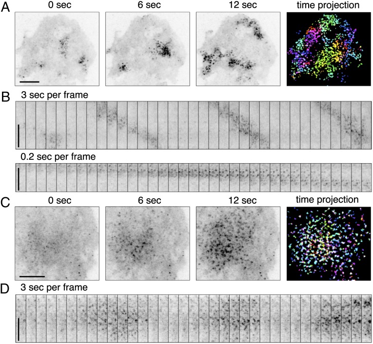
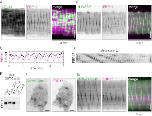
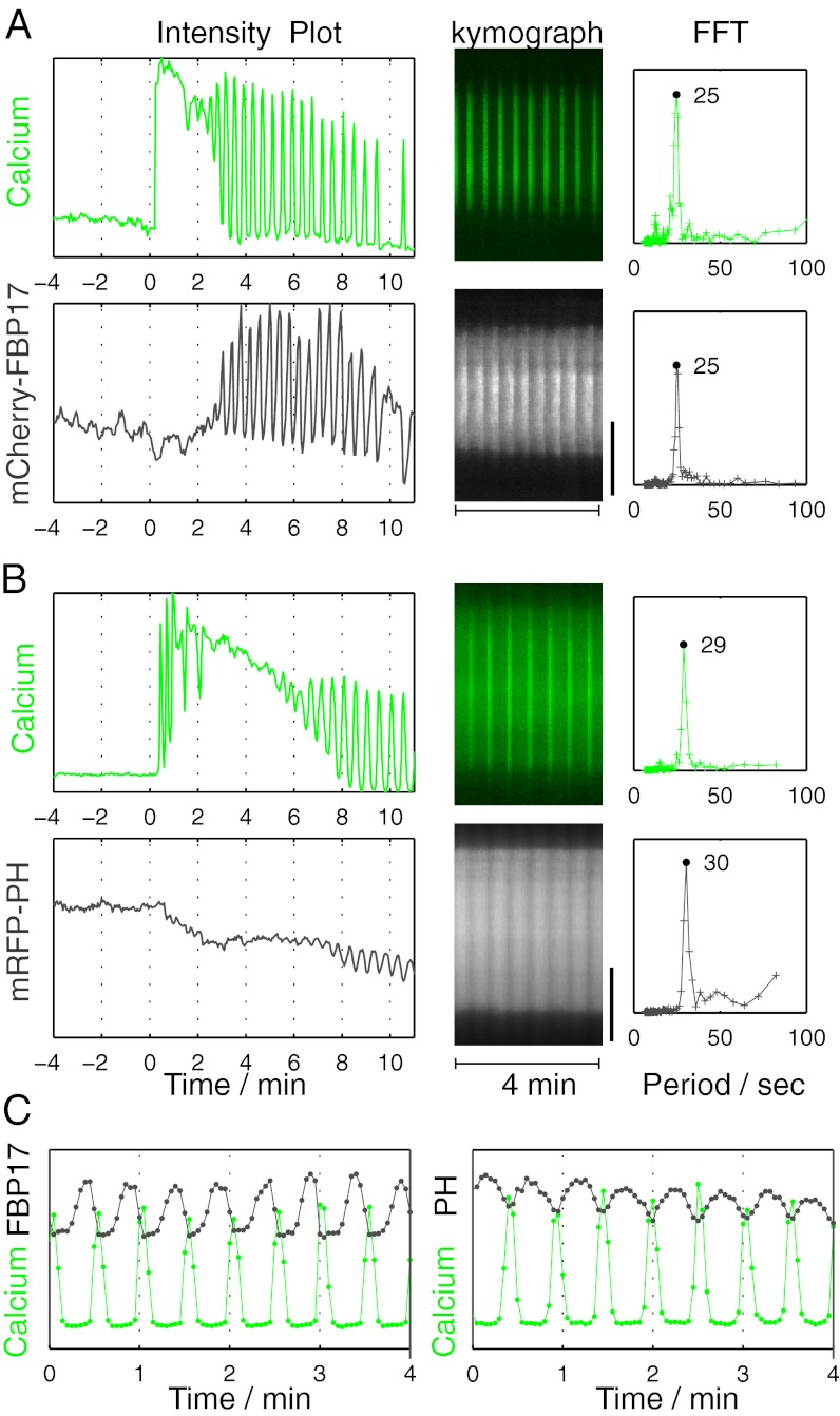
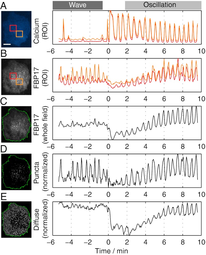
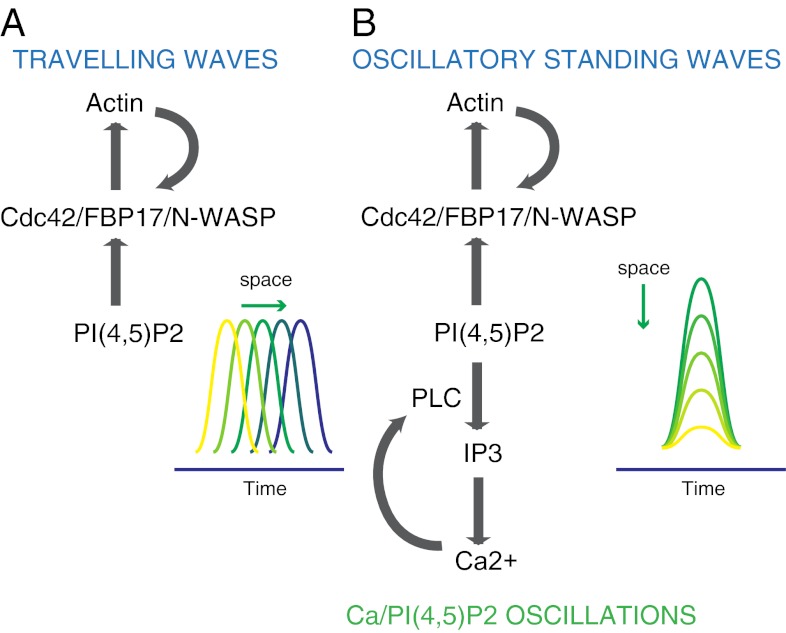
Dynamic spatial patterns of signaling factors or macromolecular assemblies in the form of oscillations or traveling waves have emerged as important themes in cell physiology. Feedback mechanisms underlying these processes and their modulation by signaling events and reciprocal cross-talks remain poorly understood. Here we show that antigen stimulation of mast cells triggers cyclic changes in the concentration of actin regulatory proteins and actin in the cell cortex that can be manifested in either spatial pattern. Recruitment of FBP17 and active Cdc42 at the plasma membrane, leading to actin polymerization, are involved in both events, whereas calcium oscillations, which correlate with global fluctuations of plasma membrane PI(4,5)P(2), are tightly linked to standing oscillations and counteract wave propagation. These findings demonstrate the occurrence of a calcium-independent oscillator that controls the collective dynamics of factors linking the actin cytoskeleton to the plasma membrane. Coupling between this oscillator and the one underlying global plasma membrane PI(4,5)P2 and calcium oscillations spatially regulates actin dynamics, revealing an unexpected pattern-rendering mechanism underlying plastic changes occurring in the cortical region of the cell.


Conflict of interest statement

The authors declare no conflict of interest.

Figures

Fig. 1.
Fig. 1.
Traveling waves or standing oscillations of FBP17 in antigen-stimulated mast cells visualized by TIRFM. (A and B) Waves. (A) Three individual frames from a movie of mCherry-FBP17 waves (Movie S1). An inverted grayscale lookup table was used, and fluorescence is shown in black. (Far Right) Color image showing superimposition of fluorescence signal from 10 different frames (3-s intervals) of the cell on the Far Left, where bright puncta in the same frame are shown in the same color. (B) Montage of 35 frames (3 s or 0.2-s intervals) of a small region of the cell showing that wave propagation is through sequential formation and disassembly of puncta. (C and D) Oscillations. (C) Three individual frames from a movie of a mCherry-FBP17 oscillation. (Far Right) Color image showing superimposition of fluorescence signal from 10 different frames (3-s intervals) of the cell shown on the Far Left, with color-coding based on time as in A. (D) Montage of 35 frames (3-s intervals) of a small region of the cell showing the cyclic assembly of mCherry-FBP17 puncta at the cell cortex. (Scale bar: 10 μm.)
Fig. 2.
Fig. 2.
Stimulus-evoked waves of FBP17 are synchronized with both N-WASP and active Cdc42 and require actin polymerization. (A and B) Kymographs of actin (LifeAct-GFP), N-WASP-GFP, and mCherry-FBP17 waves. (C) Relative phase-shifts of FBP17 and actin. Peaks of FBP17 preceded actin by 3–6 s. (D) Montage of sequential frames from a portion of a cell expressing mCherry-FBP17 showing that addition of latrunculin abolished waves (Movie S3). (E) FBP17 is a Cdc42 effector in vitro. Binding assay showing that purified FBP17 interacts with the purified active form of Cdc42, i.e., with GTPγS-bound Cdc42 or with a constitutively active GTP-bound Cdc42 mutant, but not to GDP-bound Cdc42. (F) Snapshots of a cell with antigen-evoked traveling waves show that the diffuse but locally concentrated accumulation of active Cdc42 at the plasma membrane (Cdc42 CBD-EGFP) coincides with clusters of FBP17 puncta. (G) Kymograph from a movie of traveling waves showing coupled dynamics of FBP17 and active Cdc42 (Movie S4). An inverted lookup table was used for grayscale images and kymographs. (Scale bar: 10 μm.)
Fig. 3.
Fig. 3.
Stimulation-dependent oscillations of FBP17 and PI(4,5)P2 and their relation to calcium oscillations. (A) (Left) Antigen stimulation produced an increase in the recruitment of mCherry-FBP17, which occurred as standing oscillations. Oscillations started after a sustained increase in cytosolic calcium (as detected by the calcium sensor GCaMP3), when calcium itself started to oscillate. (Center) Kymograph showing that oscillations not only of calcium but also of FBP17 occur synchronously throughout the cell. (Right) Power spectrum, based on the Fourier transforms (FFT), of calcium and mCherry-FBP17 oscillations indicating a single matching frequency. (B) Stimulation triggered oscillations of plasma membrane PI(4,5)P2, as imaged by mRFP-PHPLCδ. Center and Right are as in A. (C) Both FBP17 and PI(4,5)P2 were opposite in phase relative to calcium. In A and B, time 0 is the time of antigen addition. (Scale bar: 10 μm.)
Fig. 4.
Fig. 4.
Conversion of spontaneous traveling waves to global standing oscillations in response to a stimulus are coupled with the onset of calcium oscillations. (A) Robust calcium oscillations elicited by antigen stimulation. (B) Analysis of local FBP17 fluorescence in ROIs (orange and red squares in the micrograph) demonstrates that fluctuations in the two ROIs are not in phase before the stimulus (waves), but become synchronized after stimulation (oscillations; Movie S6). (C) Fluctuations of total FBP17 fluorescence intensities in the whole TIRFM field are more pronounced after stimulation. (D) Fluctuations of the punctate signal (white dots), normalized using the total punctate area, display a similar amplitude during waves and oscillations. (E) Oscillation of the diffuse FBP17 fluorescence (the fluorescence surrounding the puncta), normalized using the total diffuse pool area, only occurred during calcium oscillations and correlated with the conversion of traveling waves into standing oscillations. Time 0 is the time of antigen addition. (Scale bar: 10 μm.)
Fig. 5.
Fig. 5.
Schematic representation of the mechanisms underlying the traveling waves and standing oscillations. (A) Traveling waves involve actin-dependent positive-feedback mechanisms that are calcium independent. (B) Actin oscillations require similar molecular components as actin waves, but are coupled with calcium oscillations, likely via PI(4,5)P2 oscillations.

References

    1. Ozbudak EM, Becskei A, van Oudenaarden A. A system of counteracting feedback loops regulates Cdc42p activity during spontaneous cell polarization. Dev Cell. 2005;9(4):565–571. - PubMed
    1. Millius A, Dandekar SN, Houk AR, Weiner OD. Neutrophils establish rapid and robust WAVE complex polarity in an actin-dependent fashion. Curr Biol. 2009;19(3):253–259. - PMC - PubMed
    1. Giannone G, et al. Periodic lamellipodial contractions correlate with rearward actin waves. Cell. 2004;116(3):431–443. - PubMed
    1. Schroth-Diez B, et al. Propagating waves separate two states of actin organization in living cells. HFSP J. 2009;3(6):412–427. - PMC - PubMed
    1. Weiner OD, Marganski WA, Wu LF, Altschuler SJ, Kirschner MW. An actin-based wave generator organizes cell motility. PLoS Biol. 2007;5(9):e221. - PMC - PubMed

Publication types