. 2013 Jan 22;110(4):1303-8.
doi: 10.1073/pnas.1217203110. Epub 2013 Jan 7.

Nuclear export inhibition through covalent conjugation and hydrolysis of Leptomycin B by CRM1

Affiliations

Nuclear export inhibition through covalent conjugation and hydrolysis of Leptomycin B by CRM1

Qingxiang Sun et al. Proc Natl Acad Sci U S A. .

Abstract

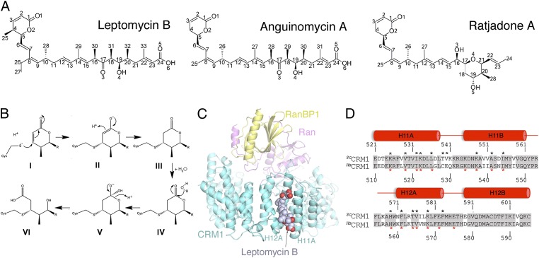
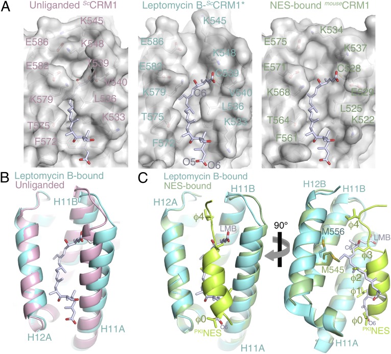
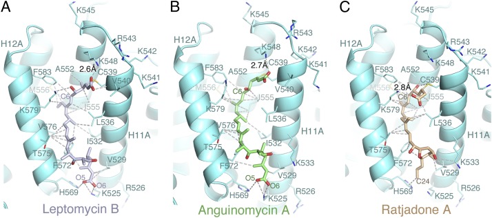
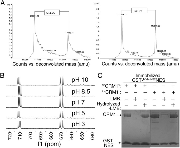
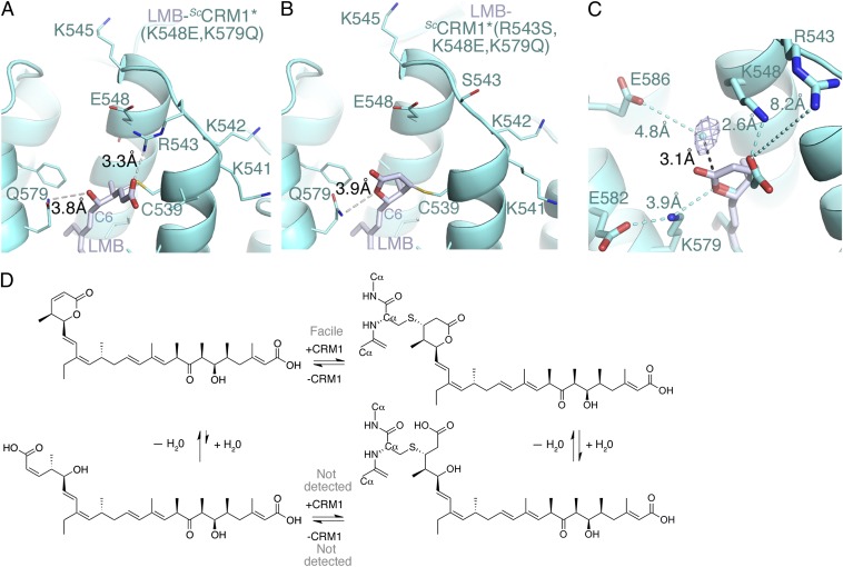
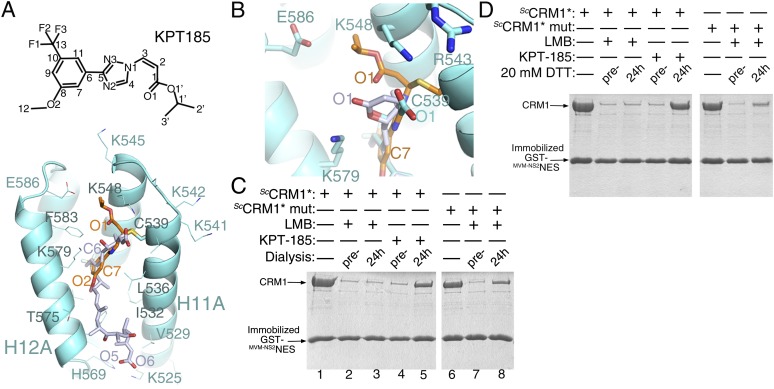
The polyketide natural product Leptomycin B inhibits nuclear export mediated by the karyopherin protein chromosomal region maintenance 1 (CRM1). Here, we present 1.8- to 2.0-Å-resolution crystal structures of CRM1 bound to Leptomycin B and related inhibitors Anguinomycin A and Ratjadone A. Structural and complementary chemical analyses reveal an unexpected mechanism of inhibition involving covalent conjugation and CRM1-mediated hydrolysis of the natural products' lactone rings. Furthermore, mutagenesis reveals the mechanism of hydrolysis by CRM1. The nuclear export signal (NES)-binding groove of CRM1 is able to drive a chemical reaction in addition to binding protein cargoes for transport through the nuclear pore complex.


Conflict of interest statement

Conflict of interest statement: Y.M.C. is a consultant for Karyopharm Therapeutics.

Figures

Fig. 1.
Fig. 1.
Chemical structures of the α,β-unsaturated lactone polyketide inhibitors and crystal structure of the LMB-bound ScCRM1*-HsRan-ScRanBP1 complex. (A) Chemical structures of inhibitors LMB, AGA, and RJA. (B) Michael addition and hydrolysis reactions of the LMB lactone. The polyketide chain of LMB is represented as R. (Upper) The deprotonated reactive cysteine of CRM1 attacks (Left) the β-alkene of the lactone (I), generating a saturated lactone that is conjugated to (Right) the cysteine (III). (Lower) A water molecule attacks the carbonyl carbon of the conjugated saturated lactone to form (Center) a tetrahedral oxyanion intermediate (V) followed by (Left) the breaking of ester bond and formation of the hydroxy acid product (VI). (C) Overall structure of LMB (space-filling representation) bound to the ternary complex of ScCRM1* (aquamarine), HsRan (magenta), and ScRanBP1 (yellow). The proteins are shown in cartoon representation. (D) Sequence alignment of the NES-binding grooves (HEAT repeats H11 and H12) of ScCRM1 and HsCRM1 (81% sequence identity). Identical residues are shaded gray. Residues that contact LMB are marked with black asterisks, and residues that contact the PKINES (Protein Data Bank ID code 3NBY) are marked with red asterisks.
Fig. 2.
Fig. 2.
Comparison of the unliganded and LMB- and NES-bound CRM1 grooves. (A) Surface representations of (Left) the unliganded NES-binding groove of ScCRM1-Ran-RanBP1 (Protein Data Bank ID code 3M1I) (15), (Center) the LMB-bound ScCRM1* groove, and (Right) the PKINES-bound HsCRM1 groove (Protein Data Bank ID code 3NBY) (10). The unliganded groove contains no LMB, which is superimposed and shown only as a reference. The PKINES peptide has been removed from the PKINES-bound HsCRM1 groove, and a superimposed LMB is shown as a reference. (B) Superposition of the unliganded (pink) and LMB-bound (aquamarine) grooves. (C) Superposition of the LMB- (aquamarine) and PKINES-bound (green) grooves.
Fig. 3.
Fig. 3.
Interactions of the inhibitors with the CRM1 grooves. Cartoon representation of the ScCRM1 grooves (aquamarine) bound with (A) LMB (light blue), (B) AGA (green), and (C) RJA (brown). Select inhibitor–CRM1 interactions (<4 Å) are shown with dashed lines.
Fig. 4.
Fig. 4.
LMB hydrolysis and CRM1 inhibition. (A) Intact mass analysis of excess ScCRM1* treated with LMB. Both samples were prepared and analyzed using the exact same protocol. The deconvoluted mass spectra show two strong protein peaks with mass shifts of 554.75 (Left) and 540.73 (Right), respectively. Although the unmodified form of ScCRM1* was detected with high mass accuracy and precision (average theoretical mass of 117,350.39 ± 0.4 Da), modified CRM1 showed a significant variation that can only be explained by molecular instability of the CRM1-bound LMB during the MS experiment. (B) 1H-NMR spectra of LMB. Lactone hydrolysis is observed only at pH 10 with ∼10% conversion to hydrolysis product after 10 min (new 1H signals at 6.50 and 6.65 ppm). (C) Pull-down inhibition assays using immobilized GST-MVM-NS2NES, ScCRM1*, HsCRM1, and LMB or hydrolyzed LMB (Coomassie-stained). LMB-modified CRM1 proteins do not bind GST-NES. Chemically hydrolyzed LMB does not inhibit ScCRM1* or HsCRM1.
Fig. 5.
Fig. 5.
Structures of LMB-bound ScCRM1 mutants and the mechanism of CRM1 inhibition. (A and B) Reaction sites in ScCRM1 double mutant ScCRM1(K548E,K579Q) and triple mutant ScCRM1(R543S,K548E,K579Q), respectively. CRM1 in aquamarine, and LMB is light blue. (C) Superposition of LMB bound to ScCRM1* and mutant ScCRM1*(R543S,K548Q,K579Q). Omit map density (1σ cutoff) is shown for the only water molecule in the reaction site. (D) A model showing the equilibria of conjugation and hydrolysis for CRM1 inhibition by LMB.
Fig. 6.
Fig. 6.
The LMB- vs. KPT-185–bound CRM1 grooves and the stability of inhibitor conjugation to CRM1. (A) Chemical structure of KPT-185 and crystal structure of the KPT-185-bound ScCRM1* groove. (B) Superposition of KPT-185– and LMB-bound ScCRM1* with LMB bound to mutant ScCRM1*(R543S,K548Q,K579Q). (C and D) ScCRM1* or mutant ScCRM1*(K541Q,K542Q,R543S,K545Q,K548Q,K579Q) were incubated with LMB or KPT-185 to achieve full CRM1 inhibition before dialysis of the samples (C) or treatment with 20 mM DTT (D) to remove excess unbound inhibitor. The extent of CRM1 inhibition was determined using pull-down inhibition assays with immobilized GST-NES, and the proteins were separated by SDS/PAGE and visualized with Coomassie staining.

References

    1. Kudo N, et al. Leptomycin B inactivates CRM1/exportin 1 by covalent modification at a cysteine residue in the central conserved region. Proc Natl Acad Sci USA. 1999;96(16):9112–9117. - PMC - PubMed
    1. Matsuyama A, et al. ORFeome cloning and global analysis of protein localization in the fission yeast Schizosaccharomyces pombe. Nat Biotechnol. 2006;24(7):841–847. - PubMed
    1. Xu D, Farmer A, Chook YM. Recognition of nuclear targeting signals by Karyopherin-β proteins. Curr Opin Struct Biol. 2010;20(6):782–790. - PMC - PubMed
    1. Kutay U, Güttinger S. Leucine-rich nuclear-export signals: Born to be weak. Trends Cell Biol. 2005;15(3):121–124. - PubMed
    1. Newlands ES, Rustin GJ, Brampton MH. Phase I trial of elactocin. Br J Cancer. 1996;74(4):648–649. - PMC - PubMed

Publication types

MeSH terms