. 2013 Apr 1;22(7):1383-94.
doi: 10.1093/hmg/dds554. Epub 2013 Jan 7.

The huntingtin N17 domain is a multifunctional CRM1 and Ran-dependent nuclear and cilial export signal

Affiliations

The huntingtin N17 domain is a multifunctional CRM1 and Ran-dependent nuclear and cilial export signal

T Maiuri et al. Hum Mol Genet. .

Abstract

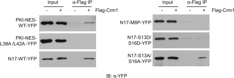
The first 17 amino acids of Huntington's disease (HD) protein, huntingtin, comprise an amphipathic alpha-helical domain that can target huntingtin to the endoplasmic reticulum (ER). N17 is phosphorylated at two serines, shown to be important for disease development in genetic mouse models, and shown to be modified by agents that reverse the disease phenotype in an HD mouse model. Here, we show that the hydrophobic face of N17 comprises a consensus CRM1/exportin-dependent nuclear export signal, and that this nuclear export activity can be affected by serine phospho-mimetic mutants. We define the precise residues that comprise this nuclear export sequence (NES) as well as the interaction of the NES, but not phospho-mimetic mutants, with the CRM1 nuclear export factor. We show that the nuclear localization of huntingtin depends upon the RanGTP/GDP gradient, and that N17 phosphorylation can also distinguish localization of endogenous huntingtin between the basal body and stalk of the primary cilium. We present a mechanism and multifunctional role for N17 in which phosphorylation of N17 not only releases huntingtin from the ER to allow nuclear entry, but also prevents nuclear export during a transient stress response event to increase the levels of nuclear huntingtin and to regulate huntingtin access to the primary cilium. Thus, N17 is a master localization signal of huntingtin that can mediate huntingtin localization between the cytoplasm, nucleus and primary cilium. This localization can be regulated by signaling, and is misregulated in HD.


Figures

Figure 1.
Figure 1.
Huntingtin N17 contains a potential conserved CRM1 NES. (A) Alignment of the N17 sequence with the LR–NES consensus sequence. (B) Representative images of N17 consensus mutants transiently expressed as YFP fusions in HEK 293 cells. Scale bar = 10 μm.
Figure 2.
Figure 2.
The huntingtin N17 NES is sensitive to leptomycin B. (A) STHdh Q7/Q7 cells stably transfected with YFP fusions of the indicated sequences were imaged after treatment with either vehicle or 10 ng/ml leptomycin B for 30 min. Scale bar = 10 μm. (B) The mean percentage nuclear fluorescence was calculated for each condition and normalized to that of the positive control (PKI-WT). Error bars = standard error of the mean for three experiments (n = 50–100 cells per condition). *P < 0.005; **P < 0.001. Scale bar = 10 μm.
Figure 3.
Figure 3.
Leptomycin B does not cause the ER UPR. The indicated cell types were treated with either 2 µg/ml tunicamycin or 10 ng/ml leptomycin B for 0–8 h prior to lysis. Equal amounts of protein were separated by SDS–PAGE and immunoblotted with anti-XBP1 antibody. *Non-specific bands.
Figure 4.
Figure 4.
N17 can bind CRM1 in the presence of RanGTP. Human HEK 293 cells were transiently transfected with the indicated YFP fusion proteins and either empty vector (−) or Flag-CRM1 and RanQ69L (+). Cell lysates were incubated with anti-Flag affinity gel (α-Flag IP). After washing, resin-associated proteins were separated by SDS–PAGE and immunoblotted with anti-YFP antibody.
Figure 5.
Figure 5.
N17 functions as an NES in the context of a huntingtin fragment. (A) STHdh Q7/Q7 cells transiently transfected with the indicated constructs were imaged after treatment with either vehicle or 10 ng/ml leptomycin B for 60 min. The mean percentage nuclear fluorescence was calculated for each condition. Error bars = standard error of the mean for three experiments (n = 50–100 cells per condition). *P < 0.005; **P < 0.001. Scale bar = 10 μm. (B) Human HEK 293 cells were transiently transfected with the indicated YFP fusion proteins and co-immunoprecipitation with Flag-CRM1 was performed as in Figure 4.
Figure 6.
Figure 6.
Disrupting the Ran gradient affects the nuclear localization of endogenous full-length huntingtin. (A) STHdh cells were transiently transfected with Flag-RERE or Flag-RanQ69L and immunofluorescence performed against the Flag epitope tag (α-flag, red) and endogenous huntingtin (α-N17, green). Scale bar = 10 μm. (B) The mean percentage nuclear fluorescence was calculated for untransfected and transfected cells. Error bars = standard error of the mean for three experiments (n = 50–100 cells per condition). ***P < 0.00002.
Figure 7.
Figure 7.
N17 phosphorylation specifies huntingtin localization between the base and stalk of the primary cilium. Co-immunofluorescence on STHdh Q7/Q7 cells was performed against acetylated tubulin to locate cilia (magenta; a, d, g, j) and either unmodified N17 (b and e) or phosphorylated N17 endogenous huntingtin (h and k). Magnified inset images show localization of unmodified N17 primarily to the cilia stalk (d–f) and phosphorylated N17 huntingtin to the basal body (j–l). White scale bars are 10 μm. Black scale bars are 2 μm. Merged magenta–green signal appears as white in (c, f, i, l).
Figure 8.
Figure 8.
Model of the role of the multifunctional N17 in huntingtin stress response. Upon ER stress or other signaling, huntingtin N17 is phosphorylated and this allows release from the ER to enter the nucleus or primary cilium via karyopherin beta2. Once in the nucleus, phospho-N17 huntingtin localizes to huntingtin chromatin-dependent nuclear puncta, where the interaction of the N17 NES with CRM1/RanGTP is occluded by phosphorylation. Upon de-phosphorylation post-stress, the NES is exposed to the CRM1/RanGTP complex and huntingtin is exported out of the nucleus. With mutant huntingtin, N17 is sterically hindered by the polyglutamine expansion, causing either poor phosphorylation of N17 to inhibit the stress response, and/or poor de-phosphorylation of nuclear mutant huntingtin resulting in nuclear accumulation and lack of proper control of the stress response. Phospho-N17 huntingtin is seen at the basal body, but not within the cilium, suggesting that a complex with CRM1 and RanGTP can mediate huntingtin export from the cilium in a mechanism identical to nuclear export.

References

    1. The Huntington's Disease Collaborative Research Group. A novel gene containing a trinucleotide repeat that is expanded and unstable on Huntington's disease chromosomes. Cell. 1993;72:971–983. doi:10.1016/0092-8674(93)90585-E. - DOI - PubMed
    1. Kim M.W., Chelliah Y., Kim S.W., Otwinowski Z., Bezprozvanny I. Secondary structure of Huntingtin amino-terminal region. Structure. 2009;17:1205–1212. doi:10.1016/j.str.2009.08.002. - DOI - PMC - PubMed
    1. Atwal R.S., Desmond C.R., Caron N., Maiuri T., Xia J., Sipione S., Truant R. Kinase inhibitors modulate huntingtin cell localization and toxicity. Nat. Chem. Biol. 2011;7:453–460. doi:10.1038/nchembio.582. - DOI - PubMed
    1. Atwal R.S., Xia J., Pinchev D., Taylor J., Epand R.M., Truant R. Huntingtin has a membrane association signal that can modulate huntingtin aggregation, nuclear entry and toxicity. Hum. Mol. Genet. 2007;16:2600–2615. doi:10.1093/hmg/ddm217. - DOI - PubMed
    1. Rockabrand E., Slepko N., Pantalone A., Nukala V.N., Kazantsev A., Marsh J.L., Sullivan P.G., Steffan J.S., Sensi S.L., Thompson L.M. The first 17 amino acids of Huntingtin modulate its sub-cellular localization, aggregation and effects on calcium homeostasis. Hum. Mol. Genet. 2007;16:61–77. doi:10.1093/hmg/ddl440. - DOI - PubMed

Publication types

MeSH terms