. 2013 Apr 1;22(7):1404-16.
doi: 10.1093/hmg/dds557. Epub 2013 Jan 7.

A new locus for X-linked dominant Charcot-Marie-Tooth disease (CMTX6) is caused by mutations in the pyruvate dehydrogenase kinase isoenzyme 3 (PDK3) gene

Affiliations

A new locus for X-linked dominant Charcot-Marie-Tooth disease (CMTX6) is caused by mutations in the pyruvate dehydrogenase kinase isoenzyme 3 (PDK3) gene

Marina L Kennerson et al. Hum Mol Genet. .

Abstract

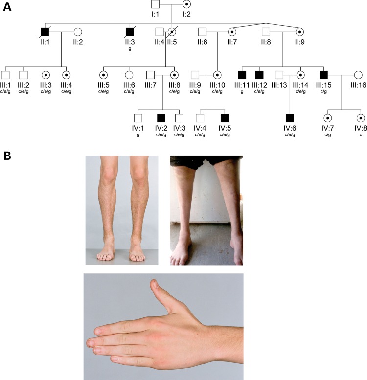
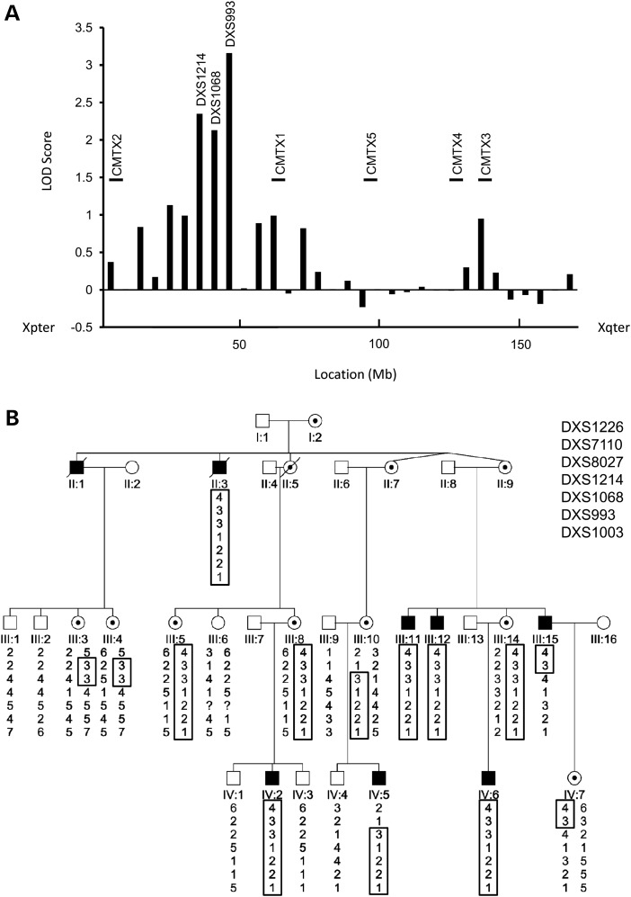
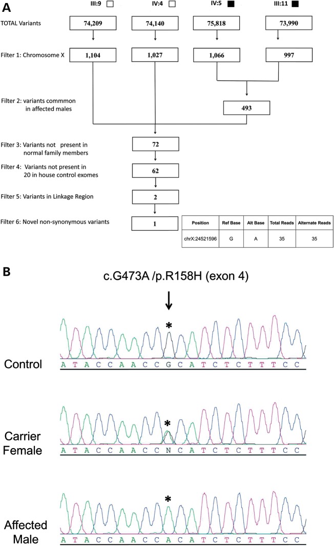
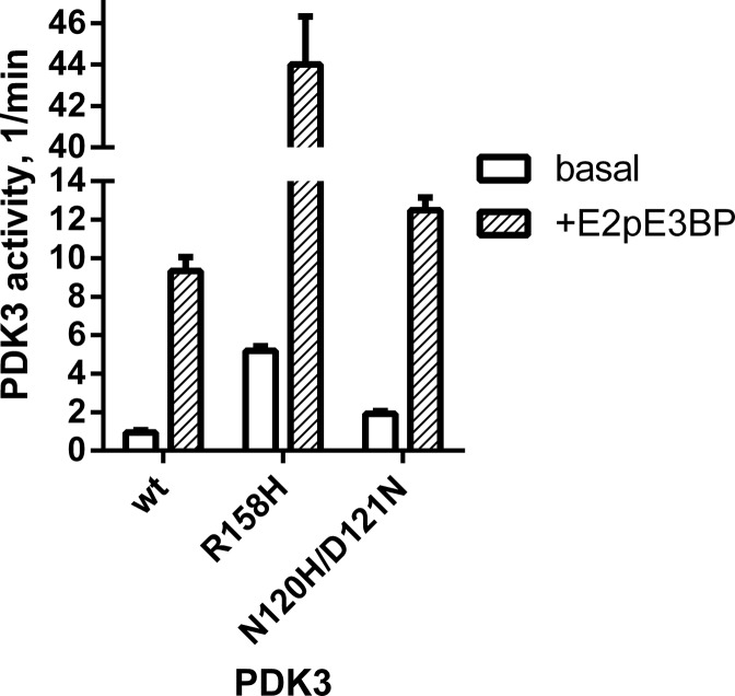
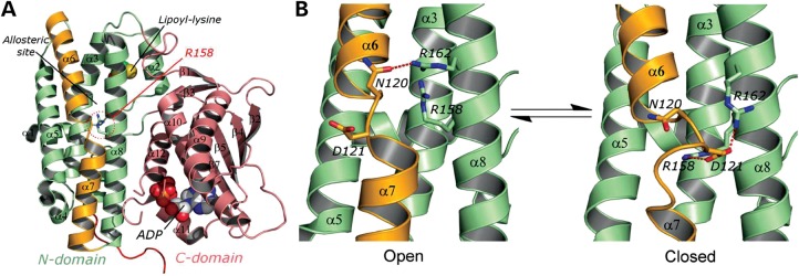
Hereditary motor and sensory disorders of the peripheral nerve form one of the most common groups of human genetic diseases collectively called Charcot-Marie-Tooth (CMT) neuropathy. Using linkage analysis in a three generation kindred, we have mapped a new locus for X-linked dominant CMT to chromosome Xp22.11. A microsatellite scan of the X chromosome established significant linkage to several markers including DXS993 (Zmax = 3.16; θ = 0.05). Extended haplotype analysis refined the linkage region to a 1.43-Mb interval flanked by markers DXS7110 and DXS8027. Whole exome sequencing identified a missense mutation c.G473A (p.R158H) in the pyruvate dehydrogenase kinase isoenzyme 3 (PDK3) gene. The change localized within the 1.43-Mb linkage interval, segregated with the affected phenotype and was excluded in ethnically matched control chromosomes. PDK3 is one of the four isoenzymes regulating the pyruvate dehydrogenase complex (PDC), by reversible phosphorylation, and is a nuclear-coded protein located in the mitochondrial matrix. PDC catalyzes the oxidative decarboxylation of pyruvate to acetyl CoA and is a key enzyme linking glycolysis to the energy-producing Krebs cycle and lipogenic pathways. We found that the R158H mutation confers enzyme hyperactivity and binds with stronger affinity than the wild-type to the inner-lipoyl (L2) domain of the E2p chain of PDC. Our findings suggest a reduced pyruvate flux due to R158H mutant PDK3-mediated hyper-phosphorylation of the PDC as the underlying pathogenic cause of peripheral neuropathy. The results highlight an important causative link between peripheral nerve degeneration and an essential bioenergetic or biosynthetic pathway required for the maintenance of peripheral nerves.


Figures

Figure 1.
Figure 1.
(A) Pedigree structure of X-linked (CMTX6) kindred. Squares indicate males, circles indicate females. Solid symbols denote affected individuals and open symbols denote unaffected family members. Internal dots indicate obligate gene carriers. Diagonal lines through symbols denote deceased individuals. The letters below individual identifiers denote the following: ‘c’ clinical examination; ‘g’ genotype examination; ‘c/e’ clinical and electrophysiological examination; ‘c/e/g’ clinical, electrophysiological and genotype examination. (B) Clinical findings in X-linked CMT (CMTX6). Affected male IV:2 aged 15 years, with mild lower limb muscle wasting (top left panel), and mild wasting of the hand intrinsic muscles (bottom panel). Affected male III:15 aged 49 years, with severe muscle wasting below the knees, and pes cavus (top right panel).
Figure 2.
Figure 2.
(A) Maximum LOD score obtained for each marker tested in X chromosome scan plotted against the location (Mb) of each marker. Markers showing significant LOD scores (DXS1214, DXS1068 and DXS993) are shown. The location axis is orientated from Xpter (left) to Xqter (right). The known CMTX loci are shown in relation to their location along the X chromosome. (B) Extended haplotype analysis of markers from chromosome Xp22.11. The haplotype segregating with the disease is boxed. The markers are ordered from the telomere (top) to the centromere (bottom) of chromosome Xp. The flanking markers defining the CMTX6 1.43-Mb interval are DXS7110 and DXS8027.
Figure 3.
Figure 3.
(A) Filtering strategy applied to variants and indels detected for individuals undergoing WES. Solid squares denote affected males; open squares denote unaffected males. (B) Sequence traces for an affected male, carrier female and normal male for p.R158H. An asterisk denotes the base position resulting in the missense mutation that segregates with CMT in the family. The GenBank sequences NM_001142386 and NP_001135858.1 were used as the reference sequences for PDK3 cDNA and PDK3 protein respectively. The mutation was designated on the basis of numbering the A in the ATG translation initiation site as +1.
Figure 4.
Figure 4.
Enzyme activity of wild-type and mutant R158H PDK3 assayed with E1p as substrate. Recombinant human PDK3 with its N-terminus fused to maltose-binding protein (MBP-PDK3) were expressed and purified. Both basal activity without the E2p/E3BP core of PDC (in open bars) and E2p/E3BP core-enhanced PDK3 activity (+E2pE3BP, in shaded bar) were measured. Enzyme activity for the N120H/D121N PDK3 double mutant was assayed with E1p and measured under basal (open bars) and core enhanced (shaded bars) conditions.
Figure 5.
Figure 5.
(A) Monomeric structure of PDK3 (PDB code: 1Y8O) is shown as ribbon models (the N-terminal domain in green, the C-terminal domain in pink, and the α6/7 helix in orange); the lipoyl-lysine residue of L2 bound to the N-terminal domain and ADP bound to the C-terminal domain are shown as space-filling models. R158 residue is shown in the stick model. (B) The close-up view of the N-terminal domain of PDK3 in equilibrium between the open conformation (PDB code: 1Y8O) and the closed conformation (molecule B of PBD code: 2PNR). The α6/7 helix (in orange) is subjected to significant iterative unwinding/winding in the equilibrium. The red dash line indicates hydrogen bonding or ionic interactions.

References

    1. Fowler W.M., Jr., Abresch R.T., Koch T.R., Brewer M.L., Bowden R.K., Wanlass R.L. Employment profiles in neuromuscular diseases. Am. J. Phys. Med. Rehabil. 1997;76:26–37. - PubMed
    1. Bergoffen J., Scherer S.S., Wang S., Scott M.O., Bone L.J., Paul D.L., Chen K., Lensch M.W., Chance P.F., Fischbeck K.H. Connexin mutations in X-linked Charcot-Marie-Tooth disease. Science. 1993;262:2039–2042. - PubMed
    1. Ionasescu V.V., Trofatter J., Haines J.L., Summers A.M., Ionasescu R., Searby C. Heterogeneity in X-linked recessive Charcot-Marie-Tooth neuropathy. Am. J. Hum. Genet. 1991;48:1075–1083. - PMC - PubMed
    1. Cowchock F.S., Duckett S.W., Streletz L.J., Graziani L.J., Jackson L.G. X-linked motor-sensory neuropathy type-II with deafness and mental retardation: a new disorder. Am. J. Med. Genet. 1985;20:307–315. - PubMed
    1. Priest J.M., Fischbeck K.H., Nouri N., Keats B.J. A locus for axonal motor-sensory neuropathy with deafness and mental retardation maps to Xq24-q26. Genomics. 1995;29:409–412. - PubMed

Publication types

MeSH terms

Substances