. 2013 Mar 8;288(10):6814-25.
doi: 10.1074/jbc.M112.393363. Epub 2013 Jan 7.

The soluble interleukin-6 receptor is a mediator of hematopoietic and skeletal actions of parathyroid hormone

Affiliations

The soluble interleukin-6 receptor is a mediator of hematopoietic and skeletal actions of parathyroid hormone

Sun Wook Cho et al. J Biol Chem. .

Abstract

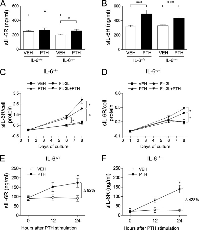
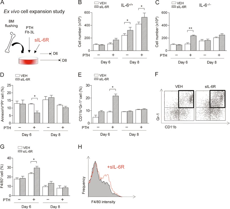
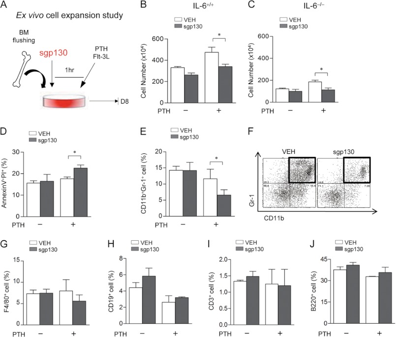
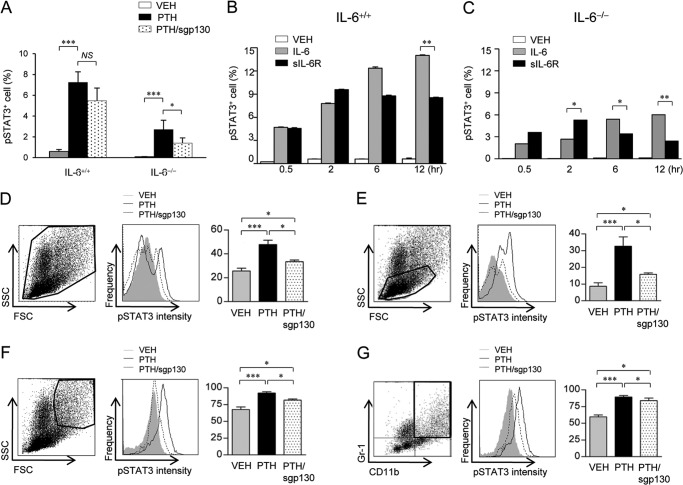
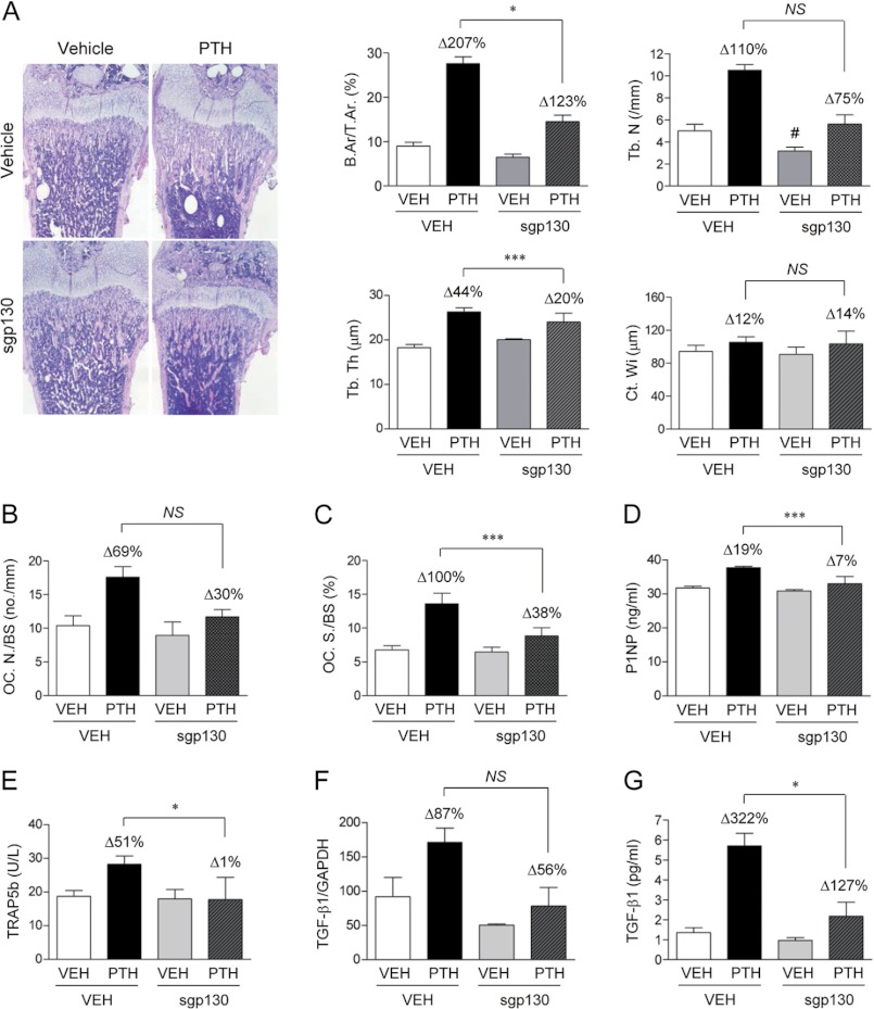
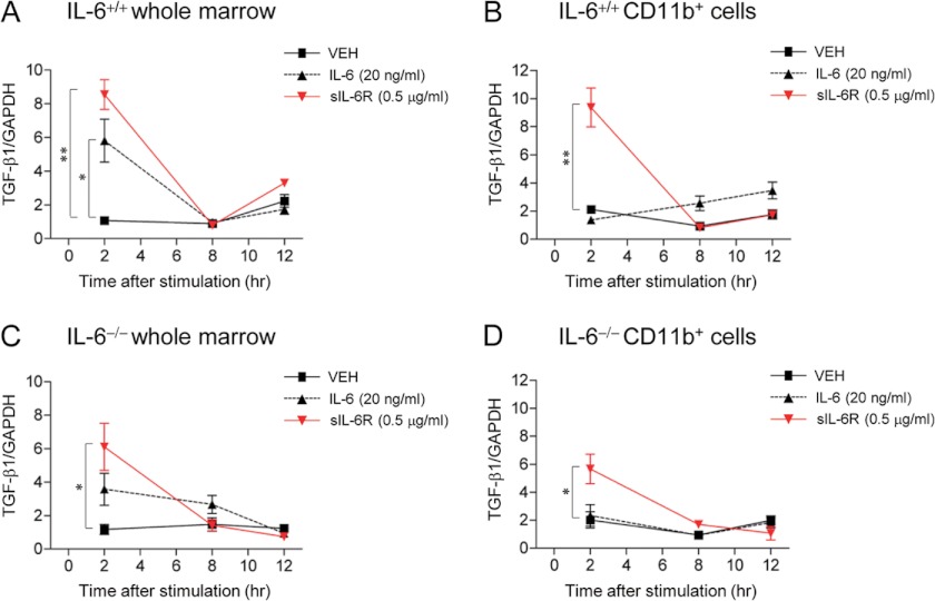
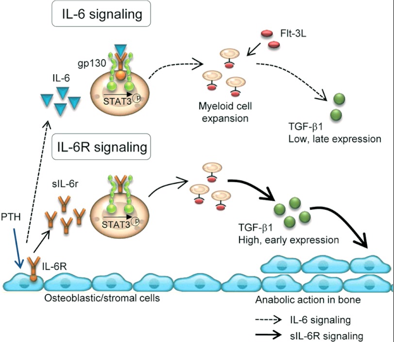
Both PTH and IL-6 signaling play pivotal roles in hematopoiesis and skeletal biology, but their interdependence is unclear. The purpose of this study was to evaluate the effect of IL-6 and soluble IL-6 receptor (sIL-6R) on hematopoietic and skeletal actions of PTH. In the bone microenvironment, PTH stimulated sIL-6R protein levels in primary osteoblast cultures in vitro and bone marrow in vivo in both IL-6(+/+) and IL-6(-/-) mice. PTH-mediated hematopoietic cell expansion was attenuated in IL-6(-/-) compared with IL-6(+/+) bone marrow, whereas sIL-6R treatment amplified PTH actions in IL-6(-/-) earlier than IL-6(+/+) marrow cultures. Blocking sIL-6R signaling with sgp130 (soluble glycoprotein 130 receptor) inhibited PTH-dependent hematopoietic cell expansion in IL-6(-/-) marrow. In the skeletal system, although intermittent PTH administration to IL-6(+/+) and IL-6(-/-) mice resulted in similar anabolic actions, blocking sIL-6R significantly attenuated PTH anabolic actions. sIL-6R showed no direct effects on osteoblast proliferation or differentiation in vitro; however, it up-regulated myeloid cell expansion and production of the mesenchymal stem cell recruiting agent, TGF-β1 in the bone marrow microenvironment. Collectively, sIL-6R demonstrated orphan function and mediated PTH anabolic actions in bone in association with support of myeloid lineage cells in the hematopoietic system.


Figures

FIGURE 1.
FIGURE 1.
Effect of PTH on sIL-6R production in bone. A and B, young (A, 3 weeks) and adult (B, 16 weeks) male and female IL-6+/+ and IL-6−/− mice were treated with PTH (50 μg/kg) or VEH for 2 weeks. sIL-6R levels were measured in bone marrow (n = 9–11/group). C and D, whole bone marrows from IL-6+/+ (C) or IL-6−/− (D) mice (4–6 weeks old, female) were seeded at 4 × 10⁶ cells/well in 24-well plates and treated once with VEH or PTH and/or Flt-3L. sIL-6R protein levels were measured from culture medium at days 1, 6, and 8. The graphs show the ratio of sIL-6R/total cell protein. Note the different y axis scales (n = 4/group). E and F, primary neonatal calvarial cells from IL-6+/+ (E) or IL-6−/− (F) mice were plated overnight in 12-well dishes at 3 × 10⁶/well followed by PTH (10 nm) stimulation in 1% serum. Culture medium was harvested 0, 12, or 24 h after PTH stimulation. Endogenous sIL-6R levels were measured by ELISA (n = 4/group). All of the numerical data are the means ± S.E. of two independent experiments. *, p < 0.05; **, p < 0.01; ***, p < 0.001.
FIGURE 2.
FIGURE 2.
Effect of sIL-6R on ex vivo hematopoietic cell expansion. A, experimental design of ex vivo cell expansion study with sIL-6R treatment. Whole bone marrows from IL-6−/− or IL-6+/+ mice (6 weeks old, female) were seeded at 1.2 × 10⁵ cells/cm2 with Flt-3L-containing medium and treated once with or without PTH. Additionally, VEH or sIL-6R (0.5 μg/ml) was co-treated in each group. Nonadherent cells were enumerated using trypan blue exclusion, and flow cytometric analysis was performed at days 6 and 8. B and C, numbers of total nonadherent cells from IL-6+/+ (B) and IL-6−/− (C) marrows. D, the percentages of annexin V+PI+ cells in IL-6−/− marrows. E, the percentages of annexin VCD11b+Gr-1+ cells from IL-6−/− marrows. F, representative dot plots of annexin VCD11b+Gr-1+ cells from PTH-treated IL-6−/− marrows at day 6. G, the percentages of annexin VF4/80+ cells from IL-6−/− marrows. H, representative histograms of annexin VF4/80+ cells from PTH-treated IL-6−/− marrows at day 6. All of the numerical data are the means ± S.E. of two independent experiments performed in triplicate. *, p < 0.05; **, p < 0.01.
FIGURE 3.
FIGURE 3.
Effect of sgp130 on ex vivo hematopoietic cell expansion. A, experimental design of ex vivo cell expansion study with sgp130 treatment. Flushed whole marrows from IL-6−/− or IL-6+/+ mice (6 weeks old, female) were pretreated with VEH or sgp130 (1 μg/ml) for 1 h and seeded at 1.2 × 10⁵ cells/cm2 with Flt-3L-containing medium. At the time of plating, each group was treated once with or without PTH. At day 8, nonadherent cells were harvested, enumerated using trypan blue exclusion, and used for flow cytometric analysis. B and C, numbers of total nonadherent cells from IL-6+/+ (B) and IL-6−/− (C) mice. D, the percentages of annexin V+PI+ cells in IL-6−/− marrows. E, the percentages of annexin VCD11b+Gr-1+ cells from IL-6−/− marrows. F, representative dot plots of annexin VCD11b+Gr-1+ cells from PTH-treated IL-6−/− marrows. G–J, plots of the percentages of annexin VF4/80+ (G), annexin VCD19+ (H), annexin VCD3+ (I), and annexin VB220+ (J) cells are shown from IL-6−/− marrows. All of the numerical data are the means ± S.E. of two independent experiments performed in triplicate. *, p < 0.05.
FIGURE 4.
FIGURE 4.
Effect of sIL-6R signaling on STAT3 phosphorylation. A, whole bone marrow cells from 4-week-old female IL-6+/+ and IL-6−/− mice were treated once ex vivo with VEH, PTH (10 nm), or PTH with 1-h pretreatment of sgp130 (1 μg/ml) for 12 h, followed by flow cytometric analysis. The graph shows the percentages of pSTAT3+ cells. B and C, whole bone marrows from 4-week-old female IL-6+/+ and IL-6−/− mice were treated once ex vivo with VEH, IL-6 (20 ng/ml), or sIL-6R (0.5 μg/ml) for 0.5, 2, 6, and 12 h. The percentages of pSTAT3+ cells from IL-6+/+ (B) and IL-6−/− (C) mice are shown. D–G, young (3 weeks) female IL-6−/− mice were treated once with VEH or sgp130 (0.3 μg) in vivo 1 h prior to treatment with PTH (50 μg/kg) or VEH. Two hours later, whole bone marrows were isolated, and flow cytometric analysis of pSTAT3+ cells was performed. D–G, analyses of pSTAT3+ cells gated in whole marrows (D), lower side scattered cells (high end SSC threshold set at 440) representing lymphocytes (E), and higher forward and side scattered cells (F, low end FSC and SSC threshold set at 420 each) representing granulocytes and myeloid cells (G, CD11bhighGr-1high). Histograms show gray (VEH), black (PTH), and dotted (PTH+sgp130) bars. All numerical data are the means ± S.E. (n = 4/group). *, p < 0.05; **, p < 0.01; ***, p < 0.001. NS, not significant; SSC, side scattered cells; FSC, forward scattered cells.
FIGURE 5.
FIGURE 5.
The role of sIL-6R transsignaling on PTH skeletal actions. sgp130 (0.3 μg) or vehicle was administrated 1 h prior to PTH (50 μg/kg) or VEH to IL-6−/− mice (3 days old) daily for 2 weeks. A, representative hematoxylin and eosin images of femur (original magnification, ×100) and plots of fractional bone area (B.Ar/T.Ar), trabecular number (Tb.N), trabecular thickness (Tb.Th), and cortical width (Ct.Wi). B and C, histomorphometric analyses of TRAP-stained bone sections including osteoclast number (B, OC.N/BS), and osteoclast surface (C, OC.S/BS). D–F, serum levels of P1NP (D), TRAP 5b (E), and mRNA (F). G, protein levels of TGF-β1 in bone marrow (n = 10–13/group). All of the numerical data are the means ± S.E. *, p < 0.05; ***, p < 0.001 corresponding to comparisons between VEH (Δ) and sgp130 (Δ); #, p < 0.05 versus VEH (first bar). NS, not significant. A Student's t test or a two-way analysis of variance was used to calculate p values.
FIGURE 6.
FIGURE 6.
Ex vivo TGF-β1 gene expression study. Whole bone marrows were harvested from 4-week-old female IL-6+/+ and IL-6−/− mice, and CD11b+ cells were sorted by magnetic-activated cell sorting. Whole marrows or CD11b+ cells were plated, and VEH, IL-6 (20 ng/ml), or sIL-6R (0.5 μg/ml) were administered for 2, 8, and 12 h ex vivo. TGF-β1 mRNA levels of were analyzed by real time PCR in IL-6+/+ whole marrow (A), IL-6+/+ myeloid (CD11b+) cells (B), IL-6−/− whole marrow (C), and IL-6−/− myeloid (D, CD11b+) cells (n = 4/group). All of the numerical data are the means ± S.E. *, p < 0.05; **, p < 0.01.
FIGURE 7.
FIGURE 7.
Working model. sIL-6R signaling mediates PTH actions in both hematopoietic and skeletal systems. PTH binds to receptors on osteoblasts/stromal cells. In response, osteoblasts produce IL-6 and sIL-6R whose signaling phosphorylates STAT3 and results in myeloid cell expansion. sIL-6R/gp130/STAT3-mediated hematopoietic cell expansion positively impacts PTH anabolic actions in bone by stimulating TGF-β1 in myeloid cells. IL-6 contributes to PTH-mediated hematopoietic cell expansion but is not essential for PTH anabolic actions in bone. sIL-6R signaling in myeloid cells results in higher and more rapid TGF-β1 expression than IL-6/gp130/STAT3 signaling. TGF-β1 has been previously shown to recruit mesenchymal stem cells to bone surfaces and support bone formation (29, 30).

References

    1. Demiralp B., Chen H. L., Koh A. J., Keller E. T., McCauley L. K. (2002) Anabolic actions of parathyroid hormone during bone growth are dependent on c-fos. Endocrinology 143, 4038–4047 - PubMed
    1. Jilka R. L., Weinstein R. S., Bellido T., Roberson P., Parfitt A. M., Manolagas S. C. (1999) Increased bone formation by prevention of osteoblast apoptosis with parathyroid hormone. J. Clin. Invest. 104, 439–446 - PMC - PubMed
    1. Bashutski J. D., Eber R. M., Kinney J. S., Benavides E., Maitra S., Braun T. M., Giannobile W. V., McCauley L. K. (2010) Teriparatide and osseous regeneration in the oral cavity. N. Engl. J. Med. 363, 2396–2405 - PMC - PubMed
    1. Chang J. K., Chang L. H., Hung S. H., Wu S. C., Lee H. Y., Lin Y. S., Chen C. H., Fu Y. C., Wang G. J., Ho M. L. (2009) Parathyroid hormone 1–34 inhibits terminal differentiation of human articular chondrocytes and osteoarthritis progression in rats. Arthritis Rheum. 60, 3049–3060 - PubMed
    1. Calvi L. M., Adams G. B., Weibrecht K. W., Weber J. M., Olson D. P., Knight M. C., Martin R. P., Schipani E., Divieti P., Bringhurst F. R., Milner L. A., Kronenberg H. M., Scadden D. T. (2003) Osteoblastic cells regulate the haematopoietic stem cell niche. Nature 425, 841–846 - PubMed

Publication types

MeSH terms