. 2013 Feb 22;288(8):5779-89.
doi: 10.1074/jbc.M112.443960. Epub 2013 Jan 6.

The Ebola virus matrix protein penetrates into the plasma membrane: a key step in viral protein 40 (VP40) oligomerization and viral egress

Affiliations

The Ebola virus matrix protein penetrates into the plasma membrane: a key step in viral protein 40 (VP40) oligomerization and viral egress

Emmanuel Adu-Gyamfi et al. J Biol Chem. .

Abstract

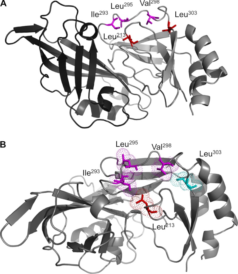
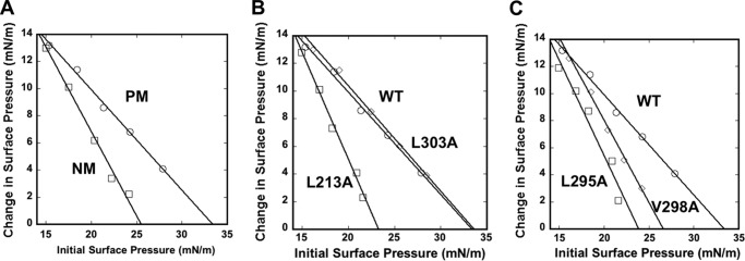
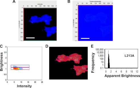
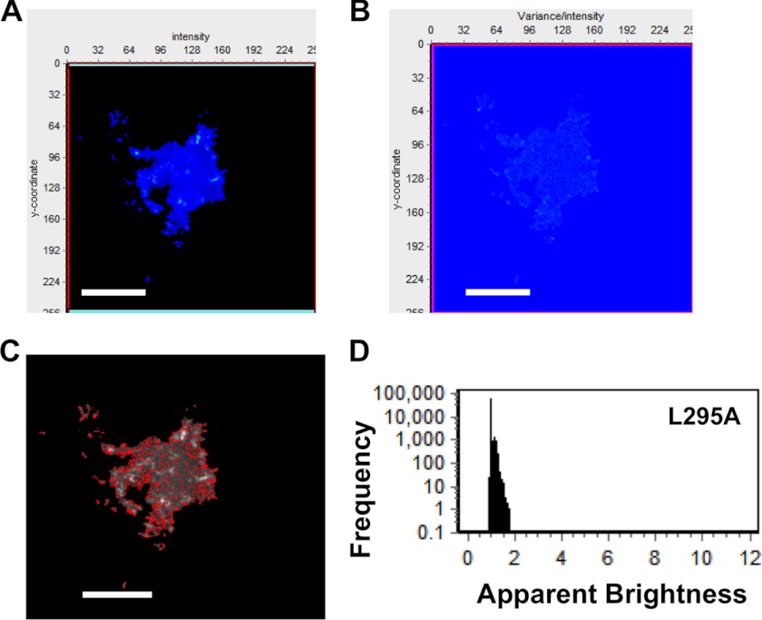
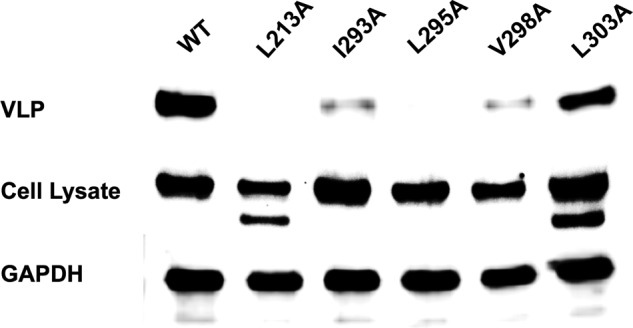
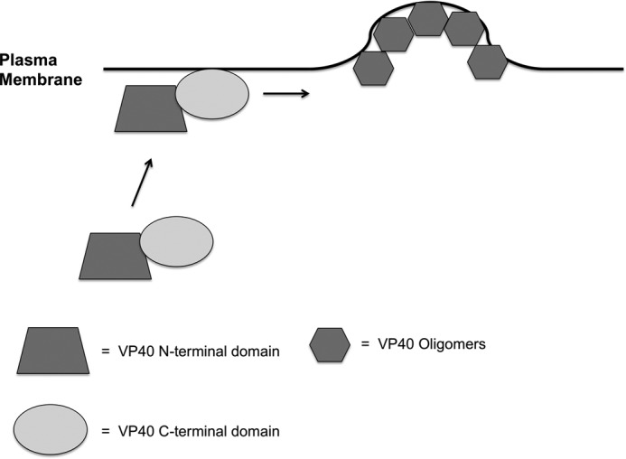
Ebola, a fatal virus in humans and non-human primates, has no Food and Drug Administration-approved vaccines or therapeutics. The virus from the Filoviridae family causes hemorrhagic fever, which rapidly progresses and in some cases has a fatality rate near 90%. The Ebola genome encodes seven genes, the most abundantly expressed of which is viral protein 40 (VP40), the major Ebola matrix protein that regulates assembly and egress of the virus. It is well established that VP40 assembles on the inner leaflet of the plasma membrane; however, the mechanistic details of plasma membrane association by VP40 are not well understood. In this study, we used an array of biophysical experiments and cellular assays along with mutagenesis of VP40 to investigate the role of membrane penetration in VP40 assembly and egress. Here we demonstrate that VP40 is able to penetrate specifically into the plasma membrane through an interface enriched in hydrophobic residues in its C-terminal domain. Mutagenesis of this hydrophobic region consisting of Leu(213), Ile(293), Leu(295), and Val(298) demonstrated that membrane penetration is critical to plasma membrane localization, VP40 oligomerization, and viral particle egress. Taken together, VP40 membrane penetration is an important step in the plasma membrane localization of the matrix protein where oligomerization and budding are defective in the absence of key hydrophobic interactions with the membrane.


Figures

FIGURE 1.
FIGURE 1.
VP40 x-ray structure depicting the N- and C-terminal domains and hydrophobic residues mutated in this study. A, the VP40 x-ray structure (Protein Data Bank code 1ES6) (40) is shown with the N-terminal domain in dark gray and the C-terminal domain in light gray. The N-terminal domain has been shown to be involved in oligomerization, and the C-terminal domain has been deemed important for membrane binding. A hydrophobic interface composed of two loops harboring Leu213, Ile293, Leu295, and Val298 was the focus of this study. All four residues were mutated to Ala. As a negative control, Leu303, adjacent but more buried than this hydrophobic interface, was also mutated to Ala. Leu303 has been shown to be part of a region in VP40 that interacts with Sec24C and was not expected to be involved in membrane penetration. B, the VP40 x-ray structure is presented looking down onto the hydrophobic interface in the C-terminal domain with key residues mutated in this study. Leu213, Ile293, Leu295, and Val298 were mutated to Ala to test their role in membrane penetration. A control residue, Leu303, is shown in cyan and was also mutated to Ala. Leu303 is just below and to the right of the hydrophobic patch we propose is involved in membrane penetration. In addition, Leu303 was a viable negative control as it has been shown to be part of a region that interacts with Sec24C. Images were generated in Mac PyMOL.
FIGURE 2.
FIGURE 2.
Monolayer penetration of VP40 and hydrophobic mutations. A, insertion of WT VP40 into a PM mimetic (POPC/POPE/POPS/POPI/cholesterol (12:35:22:9:22)) monolayer (circles) or an NM mimetic (POPC/POPE/POPS/POPI/cholesterol (61:21:4:7:7)) monolayer (squares) monitored as a function of π0. B, WT VP40 (circles), L213A (squares), and L303A (diamonds) were monitored for insertion into a PM mimetic monolayer as a function of π0. C, WT VP40 (circles), L295A (squares), and V298A (diamonds) were monitored for insertion into a PM mimetic monolayer as a function of π0. The subphase was 10 mm HEPES buffer, pH 7.4 with 0.16 m KCl for all measurements.
FIGURE 3.
FIGURE 3.
VP40 cellular localization in HEK293 cells. HEK293 cells were grown in an 8-well plate and transfected with WT VP40, L213A, I293A, L295A, V298A, or L303A DNA containing an EGFP tag. Cells were imaged after 24 h using a Zeiss LSM 710 confocal microscope with a 63× 1.4 numerical aperture oil objective. A, WT VP40 exhibits extensive PM localization and strong visual evidence of VP40-enriched particles emanating from the PM. Cells were imaged to investigate the PM localization of WT VP40 and mutations in HEK293 cells to quantify the differences between WT and mutations. B, VP40 is well established to localize and associate with the inner leaflet of the PM of mammalian cells. To demonstrate the PM localization of VP40, EGFP-VP40 was coexpressed with a robust PS sensor, the lactadherin (Lact) C2 domain (18) harboring an mCherry tag. The overlap in VP40 and lactadherin C2 signal demonstrates the predominant PM localization of VP40 as reported previously (18). PM localization was counted in WT- or mutant-expressing cells when EGFP localization was observed on the PM or as puncta associated with or structures emanating from the PM. The extent or percentage of PM localization refers to the percentage of cells in which PM localization of the EGFP tag could actually be detected with confocal microscopy. C, cellular localization of L213A was nearly undetectable in the majority of cells. D, I293A had markedly reduced PM localization in HEK293 cells. E, L295A behaved similarly to I293A with markedly reduced PM localization. F, V298A exhibited some PM localization in ∼20% of HEK293 cells, but when localization was observed it was minor (only a few diffuse spots as shown) compared with WT and the L303A control. G, L303A, a residue that is important for interactions with Sec24C, displays a similar extent of PM localization. H, a histogram was plotted to demonstrate the percentage of plasma membrane localization observed for WT and mutations in HEK293 cells. Experiments were repeated in triplicate using at least 100 cells in each experiment to determine the S.D. (error bars) as shown. *, p < 0.0002; **, p < 0.0001; NS, not significant. White scale bars, 10 μm; red scale bars, 5 μm.
FIGURE 4.
FIGURE 4.
Brightness analysis of VP40 in HEK293 cells. A, membrane protrusion sites emanating from the PM were inspected with TIRF microscopy. B, TIRF average intensity image of an HEK293 cell transfected with plasmid expressing EGFP-VP40 demonstrates sites of signal enrichment and a number of sites of membrane protrusions and viral egress. C, brightness image (variance/intensity) of the same cell demonstrates the enriched sites of VLP egress where significant EGFP signal (red) is detected. D, brightness versus intensity plot displaying monomers (brightness of 1.104) (red box), dimers (blue box), and hexamers (green box). E, brightness distribution of VP40 with selected pixels from D displaying localization of monomers (red), dimers (blue), and hexamers (green). F, frequency versus apparent brightness plot demonstrates the extensive oligomerization of VP40 at or near the PM of HEK293 cells. The apparent brightness of a monomer is 1.104, indicating the significant frequency of a monomer but extensive enrichment of oligomers up to an apparent brightness of 12. Scale bars, 18 μm.
FIGURE 5.
FIGURE 5.
Brightness versus intensity analysis of EGFP-VP40 in HEK293 cells displaying oligomers. A, brightness versus intensity plot of the HEK293 cell shown in Fig. 4 highlighting hexamers (red box), octamers (blue box), and oligomers larger than octamers (green box). B, brightness distribution of VP40 with selected pixels from A displaying hexamer (red), octamers (blue), and oligomers larger than octamers (green). Oligomeric VP40 structures are enriched on the PM and filaments protruding from the cell PM. C, brightness versus intensity plot of the HEK293 cell shown in Fig. 4 highlighting oligomers larger than octamers (green box). D, brightness distribution of VP40 with selected pixels from C displaying oligomers larger than octamers (green). Scale bars, 18 μm.
FIGURE 6.
FIGURE 6.
Brightness analysis of L213A in HEK293 cells. A, TIRF average intensity image of an HEK293 cell transfected with plasmid expressing L213A-EGFP demonstrates little localization on the PM, similar to that shown in Fig. 3. B, brightness image of the same cell demonstrates a lack of EGFP clustering or PM extension in a variance/intensity plot, indicating little oligomerization of L213A on the PM or from the premembrane zone. C, brightness versus intensity plot displaying monomers (red box) and dimers (blue box). D, brightness distribution of L213A with selected pixels from C displaying monomers (red) or dimers (blue). E, frequency versus apparent brightness plot demonstrates a lack of oligomerization of L213A at or near the PM of HEK293 cells. Scale bars, 18 μm.
FIGURE 7.
FIGURE 7.
Brightness analysis of L295A in HEK293 cells. A, TIRF average intensity image of an HEK293 cell transfected with plasmid expressing L295A-EGFP demonstrates little L295A localization on the PM. B, brightness image of the same cell demonstrates a lack of EGFP clustering or PM extensions for L295A. C, brightness distribution of VP40 with selected pixels displaying monomers (red) or dimers (blue). D, frequency versus apparent brightness plot demonstrates a lack of oligomerization of L295A at or near the PM of HEK293 cells with the majority of the L295A apparent brightness signal clustered around the monomeric value of 1.104. Scale bars, 18 μm.
FIGURE 8.
FIGURE 8.
VLP egress studies of WT VP40 and hydrophobic mutations. CHO-K1 cells were transfected with WT VP40, L213A, I293A, L295A, V298A, or L303A DNA. The cell lysate and VLPs were collected after 48 h as described earlier and subjected to Western blot with anti-EGFP. The ratio of cell lysate to VLPs was maintained for each sample with GAPDH used as a loading control for total cell density. L213A and L295A VLP intensity was not detectable in this assay, whereas I293A and V298A exhibited markedly reduced VLPs.
FIGURE 9.
FIGURE 9.
VP40 model of PM assembly, oligomerization, and egress. A model is depicted in which the C-terminal domain of VP40 is responsible for membrane penetration into the plasma membrane of the host cell. Membrane penetration of the VP40 C-terminal domain is important for VP40 oligomerization and VLP budding from the PM of host cells. In the absence of membrane penetration by VP40, VP40 oligomers and sites of VLP egress are not detectable.

References

    1. Johnson K. M., Lange J. V., Webb P. A., Murphy F. A. (1977) Isolation and partial characterisation of a new virus causing acute haemorrhagic fever in Zaire. Lancet 1, 569–571 - PubMed
    1. Elliott L. H., Kiley M. P., McCormick J. B. (1985) Descriptive analysis of Ebola virus proteins. Virology 147, 169–176 - PubMed
    1. Harty R. N. (2009) No exit: targeting the budding process to inhibit filovirus replication. Antiviral Res. 81, 189–197 - PMC - PubMed
    1. Olejnik J., Ryabchikova E., Corley R.B., Mühlberger E. (2011) Intracellular events and cell fate in filovirus infection. Viruses 3, 1501–1531 - PMC - PubMed
    1. Feldmann H., Volchkov V. E., Volchkova V. A., Klenk H. D. (1999) The glycoproteins of Marburg and Ebola virus and their potential roles in pathogenesis. Arch. Virol. Suppl. 15, 159–169 - PubMed

Publication types

MeSH terms