Review
. 2013 Jan;40(1):014301.
doi: 10.1118/1.4770279.

A review of breast tomosynthesis. Part I. The image acquisition process

Affiliations
Review

A review of breast tomosynthesis. Part I. The image acquisition process

Ioannis Sechopoulos. Med Phys. 2013 Jan.

Abstract

Mammography is a very well-established imaging modality for the early detection and diagnosis of breast cancer. However, since the introduction of digital imaging to the realm of radiology, more advanced, and especially tomographic imaging methods have been made possible. One of these methods, breast tomosynthesis, has finally been introduced to the clinic for routine everyday use, with potential to in the future replace mammography for screening for breast cancer. In this two part paper, the extensive research performed during the development of breast tomosynthesis is reviewed, with a focus on the research addressing the medical physics aspects of this imaging modality. This first paper will review the research performed on the issues relevant to the image acquisition process, including system design, optimization of geometry and technique, x-ray scatter, and radiation dose. The companion to this paper will review all other aspects of breast tomosynthesis imaging, including the reconstruction process.


Figures

Figure 1
Figure 1
Schematic of a breast tomosynthesis acquisition, in which a number of projection images is acquired of the compressed breast, while the x-ray source rotates around a center of rotation close or on the detector surface while the detector is either static or rotates, depending on the system design.
Figure 2
Figure 2
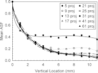
Artifact spread function from simulated DBT images acquired with a 60° angular range and varying number of projections. Reprinted with permission from I. Sechopoulos and C. Ghetti, “Optimization of the acquisition geometry in digital tomosynthesis of the breast,” Med. Phys. 36(4), 1199–1207 (2009). Copyright © 2009, American Association of Physicists in Medicine (AAPM).
Figure 3
Figure 3
Alternative x-ray source motions proposed by Zhang and Yu for acquisition of tomosynthesis projections Ref. . Reprinted with permission from J. Zhang and C. Yu, “A novel solid-angle tomosynthesis (SAT) scanning scheme,” Med. Phys. 37(8), 4186–4192 (2010). Copyright © 2010, American Association of Physicists in Medicine.
Figure 4
Figure 4
Comparison of microcalcification visibility between a standard DBT acquisition (constant exposure per projection) (left), and two different variable exposure acquisition schemes (center: central 7 projections with 4× the exposure of the 18 peripheral projections; and right: central 5 projections with 4.8× the exposure of the 20 peripheral projections), showing improved visibility for the latter. The total exposure for all three acquisitions was approximately equal. Reprinted with permission from Y.-H. Hu and W. Zhao, “The effect of angular dose distribution on the detection of microcalcifications in digital breast tomosynthesis,” Med. Phys. 38(5), 2455–2466 (2011). Copyright © 2011, American Association of Physicists in Medicine.
Figure 5
Figure 5
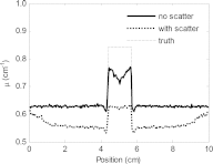
Horizontal profile through the center of a lesion in a DBT reconstructed slice showing the reduction in voxel values and contrast due to the presence of x-ray scatter. Reprinted with permission from Wu et al., “Evaluation of scatter effects on image quality for breast tomosynthesis,” Med. Phys. 36(10), 4425–4432 (2009). Copyright © 2009, American Association of Physicists in Medicine.

References

    1. NCI-funded Breast Cancer Surveillance Consortium co-operative agreement, “Performance Measures for 1,960,150 Screening Mammography Examinations from 2002 to 2006 by Age - based on BCSC data as of 2009,” November 15, 2011 [accessed April 26, 2012]; Available from: http://breastscreening.cancer.gov/data/performance/screening/2009/perf_a....
    1. Kerlikowske K., Grady D., Barclay J., Sickles E. A., and Ernster V., “Effect of age, breast density, and family history on the sensitivity of first screening mammography,” JAMA, J. Am. Med. Assoc. 276, 33–38 (1996).10.1001/jama.1996.03540010035027 - DOI - PubMed
    1. Carney P. A., Miglioretti D. L., Yankaskas B. C., Kerlikowske K., Rosenberg R., Rutter C. M., Geller B. M., Abraham L. A., Taplin S. H., Dignan M., Cutter G., and Ballard-Barbash R., “Individual and combined effects of age, breast density, and hormone replacement therapy use on the accuracy of screening mammography,” Ann. Intern Med. 138, 168–175 (2003). - PubMed
    1. Boone J. M., Nelson T. R., Lindfors K. K., and Seibert J. A., “Dedicated breast CT: Radiation dose and image quality evaluation,” Radiology 221, 657–667 (2001).10.1148/radiol.2213010334 - DOI - PubMed
    1. Ning R., Conover D. L., Chen B., Schiffhauer L., Cullinan J., Ning Y., and Robinson A. E., “Flat-panel-detector-based cone-beam volume CT breast imaging: Phantom and specimen study,” Proc. SPIE 4682, 218–227 (2002).10.1117/12.465562 - DOI

Publication types