

The central nervous system is a target of acute graft versus host disease in mice
- PMID: 23299314
- PMCID: PMC3591808
- DOI: 10.1182/blood-2012-09-456590
The central nervous system is a target of acute graft versus host disease in mice
Abstract
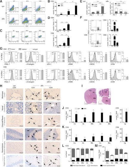
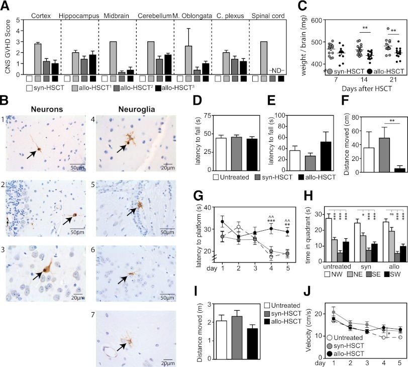
Despite significant advances in prevention and management, graft versus host disease (GVHD) is still a leading complication after allogeneic hematopoietic stem cell transplantation (allo-HSCT). Although skin, gut, liver, thymus, and lung are GVHD targets, neurological complications (NC) have also been reported following allo-HSCT. We demonstrate that the central nervous system (CNS) can be a direct target of alloreactive T cells following allo-HSCT in mice. We found significant infiltration of the CNS with donor T lymphocytes and cell death of neurons and neuroglia in allo-HSCT recipients with GVHD. We also found that allo-HSCT recipients with GVHD had deficits in spatial learning/memory and demonstrated increased anxious behavior. These findings highlight CNS sensitivity to damage caused by alloreactive donor T cells and represent the first characterization of target cell subsets and NC during GVHD. Therefore, these clinically relevant studies offer a novel and rational explanation for the well-described neurological symptoms observed after allo-HSCT.
Figures
Comment in
-
CD8-predominant T-cell CNS infiltration accompanies GVHD in primates and is improved with immunoprophylaxis.Blood. 2014 Mar 20;123(12):1967-9. doi: 10.1182/blood-2014-01-547612. Blood. 2014. PMID: 24652969 Free PMC article. No abstract available.
References
-
- Jenq RR, van den Brink MR. Allogeneic haematopoietic stem cell transplantation: individualized stem cell and immune therapy of cancer. Nat Rev Cancer. 2010;10(3):213–221. - PubMed
-
- van den Brink MR, Alpdogan O, Boyd RL. Strategies to enhance T-cell reconstitution in immunocompromised patients. Nat Rev Immunol. 2004;4(11):856–867. - PubMed
-
- Hill GR, Ferrara JL. The primacy of the gastrointestinal tract as a target organ of acute graft-versus-host disease: rationale for the use of cytokine shields in allogeneic bone marrow transplantation. Blood. 2000;95(9):2754–2759. - PubMed
-
- Antonini G, Ceschin V, Morino S, et al. Early neurologic complications following allogeneic bone marrow transplant for leukemia: a prospective study. Neurology. 1998;50(5):1441–1445. - PubMed
-
- Faraci M, Lanino E, Dini G, et al. Severe neurologic complications after hematopoietic stem cell transplantation in children. Neurology. 2002;59(12):1895–1904. - PubMed
Publication types
MeSH terms
Grants and funding
- R01 HL069929/HL/NHLBI NIH HHS/United States
- R01 CA107096/CA/NCI NIH HHS/United States
- 1R01-MH58669/MH/NIMH NIH HHS/United States
- R01-HL095075/HL/NHLBI NIH HHS/United States
- P30 CA008748/CA/NCI NIH HHS/United States
- R01-HL069929/HL/NHLBI NIH HHS/United States
- R01-CA107096/CA/NCI NIH HHS/United States
- R01 MH086883/MH/NIMH NIH HHS/United States
- R01-AI080455/AI/NIAID NIH HHS/United States
- R01 HL095075/HL/NHLBI NIH HHS/United States
- R01 MH058669/MH/NIMH NIH HHS/United States
- R01 AI080455/AI/NIAID NIH HHS/United States
- UL1 TR000457/TR/NCATS NIH HHS/United States
- 5R01-MH086883/MH/NIMH NIH HHS/United States
LinkOut - more resources
Full Text Sources
Other Literature Sources
Medical
