

Disruption of claudin-18 diminishes ovariectomy-induced bone loss in mice
- PMID: 23299504
- PMCID: PMC3602660
- DOI: 10.1152/ajpendo.00408.2012
Disruption of claudin-18 diminishes ovariectomy-induced bone loss in mice
Abstract
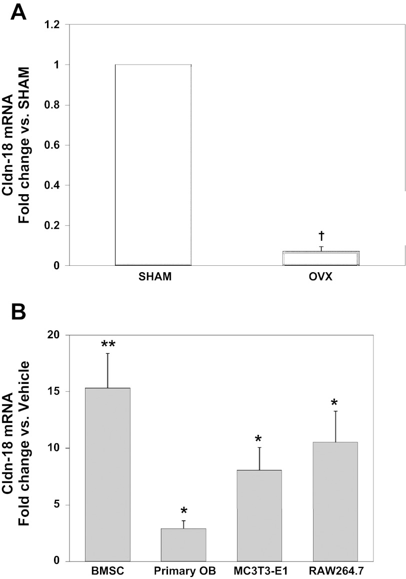
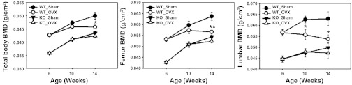
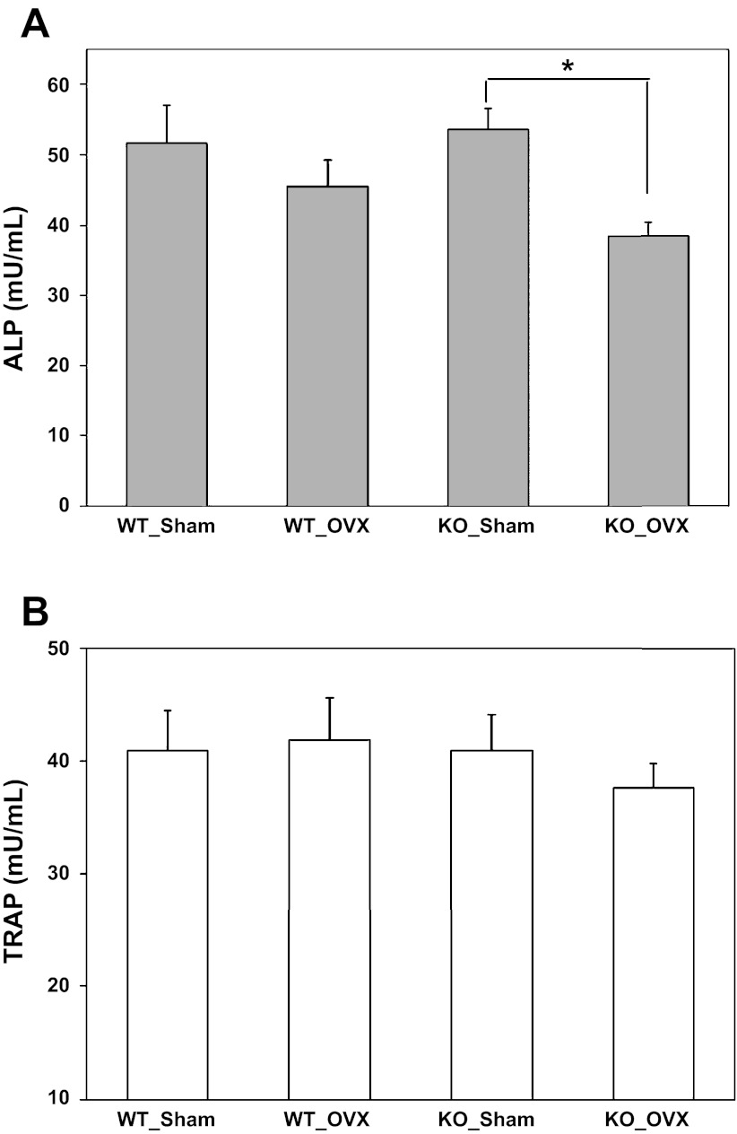
Claudin-18 (Cldn-18), a member of the tight junction family of proteins, is a negative regulator of RANKL-induced osteoclast differentiation and bone resorption (BR) in vivo. Since estrogen deficiency decreases bone mass in part by a RANKL-mediated increase in BR, we evaluated whether estrogen regulates Cldn-18 expression in bone. We found that Cldn-18 expression was reduced in the bones of estrogen deficient mice, whereas it was increased by estrogen treatment in osteoblasts and osteoclasts in vitro. We next evaluated the role of Cldn-18 in mediating estrogen-induced bone loss. Cldn-18 knockout (KO) and littermate wild-type (WT) mice were ovariectomized (OVX) or sham operated at 6 wk of age, and the skeletal phenotype was evaluated at 14 wk of age. PIXImus revealed that total body, femur, and lumbar BMD were reduced 8-13% (P < 0.05) after 8 wk of OVX compared with sham in WT mice. As expected, total body, femur, and lumbar BMD were reduced 14-21% (P < 0.05) in Cldn-18 KO sham mice compared with sham WT mice. However, ovariectomy failed to induce significant changes in BMD of total body, femur, or vertebra in the Cldn-18 KO mice. μCT analysis of the distal femur revealed that trabecular (Tb) bone volume was decreased 50% in the OVX WT mice compared with sham that was caused by a 26% decrease in Tb number and a 30% increase in Tb separation (all P < 0.05). By contrast, none of the Tb parameters were significantly different in OVX Cldn-18 KO mice compared with sham KO mice. Histomorphometric analyses at the Tb site revealed that neither osteoclast surface nor osteoclast perimeter was increased significantly as a consequence of OVX in either genotype at the time point examined. Based on our findings, we conclude that the estrogen effects on osteoclasts may in part be mediated via regulation of Cldn-18 signaling.
Figures

Similar articles
-
Emerging multifunctional roles of Claudin tight junction proteins in bone.Endocrinology. 2014 Jul;155(7):2363-76. doi: 10.1210/en.2014-1173. Epub 2014 Apr 23. Endocrinology. 2014. PMID: 24758302 Free PMC article. Review.
-
Claudin 18 is a novel negative regulator of bone resorption and osteoclast differentiation.J Bone Miner Res. 2012 Jul;27(7):1553-65. doi: 10.1002/jbmr.1600. J Bone Miner Res. 2012. PMID: 22437732 Free PMC article.
-
Effects of JSOG-6 on protection against bone loss in ovariectomized mice through regulation of osteoblast differentiation and osteoclast formation.BMC Complement Altern Med. 2014 Jun 6;14:184. doi: 10.1186/1472-6882-14-184. BMC Complement Altern Med. 2014. PMID: 24903150 Free PMC article.
-
Critical role of beta3 integrin in experimental postmenopausal osteoporosis.J Bone Miner Res. 2005 Dec;20(12):2116-23. doi: 10.1359/JBMR.050724. Epub 2005 Jul 25. J Bone Miner Res. 2005. PMID: 16294265
-
Deficiency of adiponectin protects against ovariectomy-induced osteoporosis in mice.PLoS One. 2013 Jul 2;8(7):e68497. doi: 10.1371/journal.pone.0068497. Print 2013. PLoS One. 2013. PMID: 23844209 Free PMC article.
Cited by
-
Novel Role for Claudin-11 in the Regulation of Osteoblasts via Modulation of ADAM10-Mediated Notch Signaling.J Bone Miner Res. 2019 Oct;34(10):1910-1922. doi: 10.1002/jbmr.3763. Epub 2019 Jul 26. J Bone Miner Res. 2019. PMID: 31112308 Free PMC article.
-
Emerging multifunctional roles of Claudin tight junction proteins in bone.Endocrinology. 2014 Jul;155(7):2363-76. doi: 10.1210/en.2014-1173. Epub 2014 Apr 23. Endocrinology. 2014. PMID: 24758302 Free PMC article. Review.
-
Glucose absorption in the duodenum is modulated by an estrogen receptor α-dependent regulation of glucose transporter functional expression.BMJ Open Diabetes Res Care. 2025 Jul 27;13(4):e004914. doi: 10.1136/bmjdrc-2025-004914. BMJ Open Diabetes Res Care. 2025. PMID: 40716951 Free PMC article.
-
Concurrent muscle and bone deterioration in a murine model of cancer cachexia.Physiol Rep. 2013 Nov;1(6):e00144. doi: 10.1002/phy2.144. Epub 2013 Nov 7. Physiol Rep. 2013. PMID: 24400146 Free PMC article.
-
Estrogen regulates duodenal calcium absorption and improves postmenopausal osteoporosis by the effect of ERβ on PMCA1b.Sci Rep. 2025 May 8;15(1):16053. doi: 10.1038/s41598-025-00605-2. Sci Rep. 2025. PMID: 40341576 Free PMC article.
References
-
- Bord S, Ireland DC, Beavan SR, Compston JE. The effects of estrogen on osteoprotegerin, RANKL, and estrogen receptor expression in human osteoblasts. Bone 32: 136–141, 2003 - PubMed
-
- Burek M, Arias-Loza PA, Roewer N, Forster CY. Claudin-5 as a novel estrogen target in vascular endothelium. Arterioscler Thromb Vasc Biol 30: 298–304, 2010 - PubMed
-
- Eriksen EF, Colvard DS, Berg NJ, Graham ML, Mann KG, Spelsberg TC, Riggs BL. Evidence of estrogen receptors in normal human osteoblast-like cells. Science 241: 84–86, 1988 - PubMed
-
- Furuse M. Knockout animals and natural mutations as experimental and diagnostic tool for studying tight junction functions in vivo. Biochim Biophys Acta 1788: 813–819, 2009 - PubMed
Publication types
MeSH terms
Substances
Grants and funding
LinkOut - more resources
Full Text Sources
Other Literature Sources
Molecular Biology Databases
Research Materials