

Hand-Schüller-Christian disease and Erdheim-Chester disease: coexistence and discrepancy
- PMID: 23299772
- PMCID: PMC3556249
- DOI: 10.1634/theoncologist.2012-0234
Hand-Schüller-Christian disease and Erdheim-Chester disease: coexistence and discrepancy
Abstract
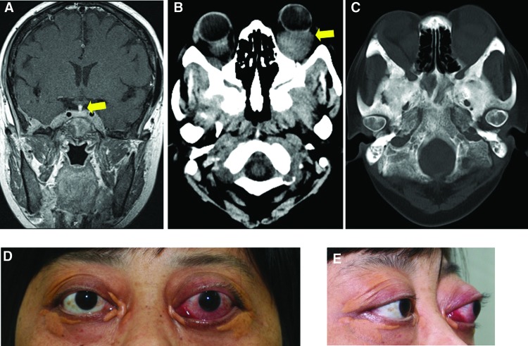
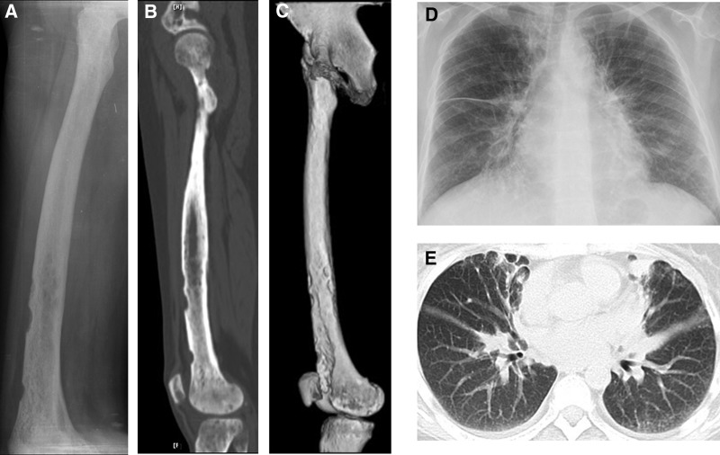
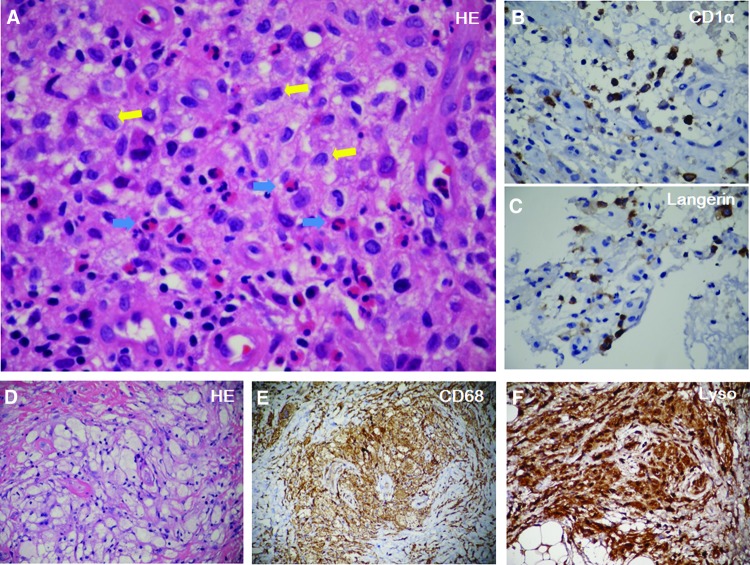
Langerhans cell histiocytosis (LCH) and Erdheim-Chester disease (ECD) share similar clinical features and mechanisms. In very rare circumstances, the two diseases coexist in the same patient. Here we report such a patient, who was first diagnosed with Hand-Schüller-Christian disease (HSC), a type of LCH. Several years later, the patient presented with severe exophthalmos and osteosclerosis on radiograph. New biopsy revealed ECD. We also analyze 54 cases of LCH and 6 cases of ECD diagnosed in our hospital, as well as their progression during a follow-up period of 8 years. In five cases of HSC (9.3% of LCH), a triad of central diabetes insipidus, hyperprolactinemia, and pituitary stalk thickening on magnetic resonance imaging (MRI) preceded the typical bone lesions by 4-9 years. In addition, LCH was featured as elevated plasma alkaline phosphatase (ALP), which was normal in ECD. Combined with a literature review, several features are summarized to differentiate ECD from HSC. In patients with diabetes insipidus, concomitant hyperprolactinemia and pituitary stalk thickening on MRI indicate a possible HSC. Additionally, if osteosclerosis is observed in a patient with LCH, the coexistence of ECD should be considered.
Conflict of interest statement
Disclosures of potential conflicts of interest may be found at the end of this article.
Figures
Comment in
-
A tale of two histiocytic disorders.Oncologist. 2013;18(1):2-4. doi: 10.1634/theoncologist.2012-0440. Epub 2013 Jan 8. Oncologist. 2013. PMID: 23299771 Free PMC article.
References
-
- Broadbent V, Gadner H, Komp DM, et al. Histiocytosis syndromes in children: II. Approach to the clinical and laboratory evaluation of children with langerhans cell histiocytosis. Med Pediatr Oncol. 1989;17:492–495. - PubMed
-
- Wilejto M, Abla O. Langerhans cell histiocytosis and Erdheim-Chester disease. Curr Opin Rheumatol. 2012;24:90–96. - PubMed
-
- Orii T, Takeda H, Kawata S, et al. Differential immunophenotypic analysis of dendritic cell tumours. J Clin Pathol. 2010;63:497–503. - PubMed
-
- Singhi AD, Montgomery EA. Gastrointestinal tract Langerhans cell histiocytosis: A clinicopathologic study of 12 patients. Am J Surg Pathol. 2011;35:305–310. - PubMed
Publication types
MeSH terms
Substances
LinkOut - more resources
Full Text Sources
Other Literature Sources
