. 2013 Jan 8;2(1):e67.
doi: 10.1038/mtna.2012.59.

In vivo SELEX for Identification of Brain-penetrating Aptamers

Affiliations

In vivo SELEX for Identification of Brain-penetrating Aptamers

Congsheng Cheng et al. Mol Ther Nucleic Acids. .

Abstract

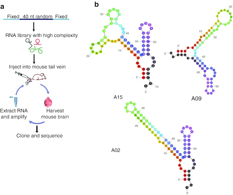
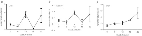
The physiological barriers of the brain impair drug delivery for treatment of many neurological disorders. One delivery approach that has not been investigated for their ability to penetrate the brain is RNA-based aptamers. These molecules can impart delivery to peripheral tissues and circulating immune cells, where they act as ligand mimics or can be modified to carry payloads. We developed a library of aptamers and an in vivo evolution protocol to determine whether specific aptamers could be identified that would home to the brain after injection into the peripheral vasculature. Unlike biopanning with recombinant bacteriophage libraries, we found that the aptamer library employed here required more than 15 rounds of in vivo selection for convergence to specific sequences. The aptamer species identified through this approach bound to brain capillary endothelia and penetrated into the parenchyma. The methods described may find general utility for targeting various payloads to the brain.Molecular Therapy - Nucleic Acids (2013) 2, e67; doi:10.1038/mtna.2012.59; published online 8 January 2013.


Figures

Figure 1
Figure 1
In vivo SELEX for aptamers. (a) Schema for SELEX strategy. An RNA library consisting of a 40 nt random region flanked 5′ by a 32 bp left arm containing a T7 transcription start, and 3′ by a 16 bp for PCR amplification was generated, and then RNAs transcribed in the presence of 2′-fluoro nucleosides to enhance nuclease resistance. Purified RNAs were peripherally injected into three C57BL/6 mice and circulated for 1 hour (rounds 1–8) or 3 hours (rounds 9–22). Mice were perfused with DPBS, and RNAs were extracted from whole brain. Recovered RNAs were treated with RNAse A and DNase I, and then converted to double-stranded DNA by RT-PCR. Purified DNAs were transcribed into 2′-F RNA, and full-length RNA was purified for the subsequent round of panning. (b) Predicted secondary structures of aptamers using Mfold (http://mfold.rna.albany.edu/?q=mfold/RNA-Folding-Form). DPBS, Dulbecco's phosphate-buffered saline; nt, nucleotide; RT-PCR, reverse transcription PCR; SELEX, Systematic Evolution of Ligands by EXponential enrichment.
Figure 2
Figure 2
Assessment for aptamer enrichment. RNA (1 nmol) from the starting library (R0), round 6 (R6), round 12 (R12), round 18 (R18), or round 22 (R22) were injected into C57BL/6 mice (three mice/library). Three hours post-injection, the mice were perfused and total RNAs from the (a) liver, (b) kidney or (c) brain were extracted and the relative abundance of RNA library in these organs was quantified by qRT-PCR. Data are normalized to 5S RNA and are presented as mean ± SD. qRT-PCR, quantitative reverse transcription-PCR; SELEX, Systematic Evolution of Ligands by EXponential enrichment.
Figure 3
Figure 3
The A15 aptamer shows enhanced specificity to brain. (a) Data represent the percentage of the denoted aptamer present after sequencing of clones from SELEX. (b) Data represents the percentage of the denoted aptamer found in the reverse screening assay. For this, equal amounts of selected RNA motifs (A02, A09 or A15, see Table 1) and scrambled A15 (SCAP) were mixed, and injected into mice. RNA was extracted from the brain, reverse transcribed, cloned, and colonies were sequenced. The frequency of the sequences representing each aptamer is shown. (c) Equal amounts of aptamers (SCAP and A15) were peripherally injected in mice (n = 3–4) and after 4 hours, total RNA was isolated. Aptamer quantitation in brain, kidney, and liver was done using one-step qRT-PCR (5 µg of DNase-treated input RNA) by including 18S RNA as loading control. (d) Quantification of A15 aptamer in capillary versus parenchymal tissues after peripheral injection. Mouse brain capillaries were clarified from brain parenchymal fractions by density centrifugation, and the purity of the capillary fraction was evaluated by assessing the endothelial cell-specific markers Tie-2, Claudin-5, and Occludin. mRNA levels were normalized to GAPDH. Data are expressed as means ± SD (*P < 0.05, Student's t-test; n = 3). (e) qRT-PCR to evaluate aptamer abundance in the indicated fractions. Data are presented as means ± SD using ANOVA and Bonferroni post hoc for significance (*P < 0.05; n = 3 mice per group). ANOVA, analysis of variance; GAPDH, glyceraldehyde 3-phosphate dehydrogenase; qRT-PCR, quantitative reverse transcription-PCR; SELEX, Systematic Evolution of Ligands by EXponential enrichment.
Figure 4
Figure 4
Aptamer biodistribution after peripheral injection. Representative in situ hybridization images from cerebral cortex (Cx), striatum (Str), hippocampus (Hp), and cerebellum (Cb) of mice, collected 3 hours after carotid artery injection with either scrambled (left) or A15 (right) aptamer. The slides were lightly counterstained with methylene green (cell nuclei; light blue). Purple precipitate denotes aptamer hybridization signal (note difference in intensity of aptamer signal between right panels and left panels). Arrows denote regions magnified in insets (lower right, all images) for better visualization of aptamer signal. Sections are representative of those from three different mice/group. Bar = 100 µm and applies to all images. Inset magnification ×5.
Figure 5
Figure 5
SELEX-derived aptamers show improved binding and uptake into mouse brain endothelial cells in vitro. (a) Flow cytometry to monitor aptamer binding and uptake on bEnd.3 cells. The bEnd.3 cells were treated with the denoted aptamers at 4 or 37 °C. The panels show that A15 binding is enhanced over SCAP (left panel), and that there is extensive uptake at 37 °C relative to SCAP (right panel). (b) Representative confocal images of bEnd.3 cells after incubation with 500 nmol/l SA-PE–conjugated A15 or SCAP, showing cellular uptake with A15, but not SCAP. Green, Alexa-488 labeled wheat germ agglutinin for cell membrane labeling; blue, DAPI to stain nuclei; red, SA-PE–conjugated aptamer. Bar = 20 µm. (c) Quantitation of aptamer internalization into brain endothelia cells. Mouse brain endothelial cells were blocked with 50 µg/ml yeast tRNA and 1 µg/µl poly d(I:C) for 30 minutes, and treated with 1 µmol/l A15 or SCAP for 2 hours at 37 °C. Cells were treated with 0.02 U/µl of Riboshredder to degrade residual RNAs on the cell surface or in the media. Internalized RNA was quantified by qPCR and normalized to GAPDH. The results are presented as means ± SD. Statistical significance was done using ANOVA and Bonferroni post hoc (*P < 0.05; n = 6 per group). ANOVA, analysis of variance; DAPI, 4′,6-diamidino-2-phenylindole; GAPDH, glyceraldehyde 3-phosphate dehydrogenase; qPCR, quantitative PCR; SA-PE, streptavidin-phycoerythrin; tRNA, transfer RNA.

References

    1. de Boer AG., and, Gaillard PJ. Drug targeting to the brain. Annu Rev Pharmacol Toxicol. 2007;47:323–355. - PubMed
    1. Chen Y., and, Liu L. Modern methods for delivery of drugs across the blood-brain barrier. Adv Drug Deliv Rev. 2012;64:640–665. - PubMed
    1. Pardridge WM. Blood-brain barrier biology and methodology. J Neurovirol. 1999;5:556–569. - PubMed
    1. Pavan B, Dalpiaz A, Ciliberti N, Biondi C, Manfredini S., and, Vertuani S. Progress in drug delivery to the central nervous system by the prodrug approach. Molecules. 2008;13:1035–1065. - PMC - PubMed
    1. Davidson BL., and, Breakefield XO. Viral vectors for gene delivery to the nervous system. Nat Rev Neurosci. 2003;4:353–364. - PubMed