. 2013 Feb 6;32(3):473-86.
doi: 10.1038/emboj.2012.342. Epub 2013 Jan 8.

Chtop is a component of the dynamic TREX mRNA export complex

Affiliations

Chtop is a component of the dynamic TREX mRNA export complex

Chung-Te Chang et al. EMBO J. .

Abstract

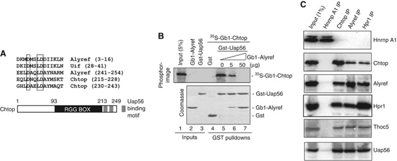
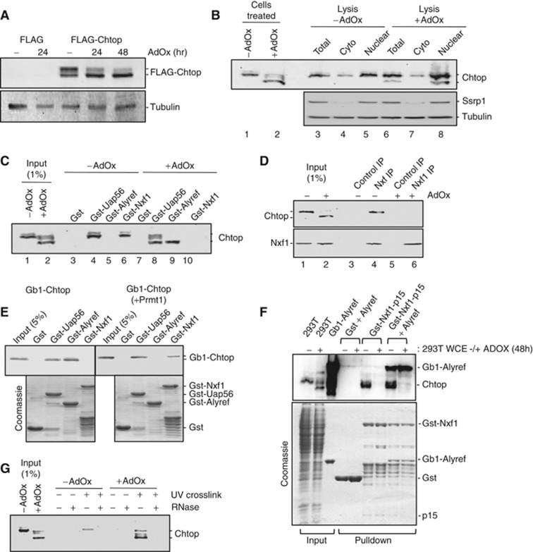
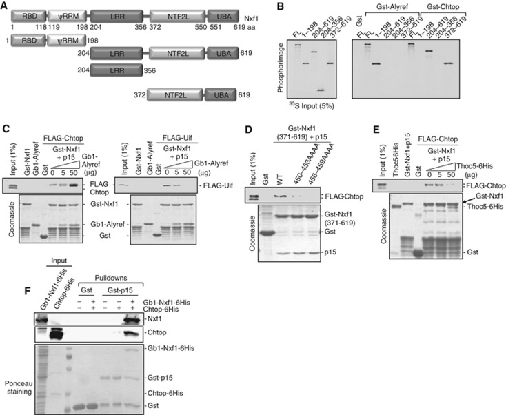
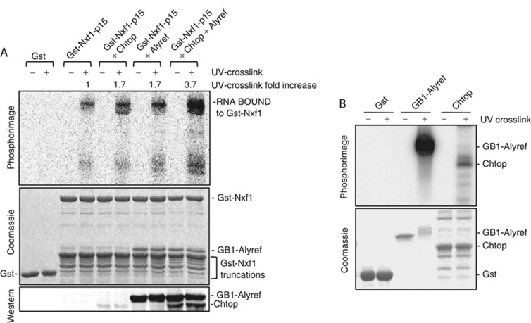
The TREX complex couples nuclear pre-mRNA processing with mRNA export and contains multiple protein components, including Uap56, Alyref, Cip29 and the multi-subunit THO complex. Here, we have identified Chtop as a novel TREX component. We show that both Chtop and Alyref activate the ATPase and RNA helicase activities of Uap56 and that Uap56 functions to recruit both Alyref and Chtop onto mRNA. As observed with the THO complex subunit Thoc5, Chtop binds to the NTF2-like domain of Nxf1, and this interaction requires arginine methylation of Chtop. Using RNAi, we show that co-knockdown of Alyref and Chtop results in a potent mRNA export block. Chtop binds to Uap56 in a mutually exclusive manner with Alyref, and Chtop binds to Nxf1 in a mutually exclusive manner with Thoc5. However, Chtop, Thoc5 and Nxf1 exist in a single complex in vivo. Together, our data indicate that TREX and Nxf1 undergo dynamic remodelling, driven by the ATPase cycle of Uap56 and post-translational modifications of Chtop.


Conflict of interest statement

The authors declare that they have no conflict of interest.

Figures

Figure 1
Figure 1
Chtop interacts with Uap56 and TREX. (A) Alignment of the Uap56-binding motifs (UBMs) present in Alyref, Uif and Chtop proteins. (B) Pulldown competition assay with Gst-Uap56 complexed with 35S-Gb1-Chtop and increasing amounts of Gb1-Alyref. Proteins were detected by Coomassie staining and Phosphorimaging. (C) Co-IP of Chtop with TREX subunits using RNAse A-treated 293T cell extract. HnrnpA1, Chtop, Alyref and Hpr1 were IPed using antibodies to the endogenous proteins and co-immunoprecipitating proteins were detected by western blotting with the indicated antibodies. Source data for this figure is available on the online supplementary information page.
Figure 2
Figure 2
Chtop activates the ATPase and helicase activity of Uap56. (A) ATPase activities for purified Uap56 in the presence of Alyref, Chtop, Cip29, RNA and ATP as indicated. Values are the average from three independent assays and error bars represent the standard deviation. Values are shown relative to the values observed for Uap56+ATP+RNA. (B) The helicase assay was performed at 37°C for 15 min with 1 μM Uap56, 2 μM Alyref, Chtop, Cip29 or Magoh. (C) In vitro protein:RNA UV crosslinking assay. 2 μg Gst-Uap56, Gb1-Alyref and Magoh purified from E. coli and Chtop purified from Baculovirus-infected cells were UV crosslinked with a 32P-radiolabelled RNA oligonucleotide in the presence or absence of ATP as indicated. Resulting complexes were analysed by SDS–PAGE and visualised by Coomassie blue staining and Phosphorimaging. (D) In vitro reconstitution of Uap56-RNA-Alyref complexes. Purified Uap56 expressed in E. coli was first incubated with continuously 32P-radiolabelled RNA and non-hydrolysable ATP. Recombinant Gst or Gst-Alyref was added to the reactions when indicated. Bound RNA was crosslinked (+) or not (−) to the proteins by UV irradiation, treated with RNAse A, and the resulting Uap56-RNA-Alyref complexes were purified using glutathione Sepharose. Eluted complexes were analysed by SDS–PAGE and visualised by Coomassie blue (left panel) and Phosphorimaging (right panel). (E) mRNP capture assay. Poly(A)+ RNA from stable Flp-In 293 cells, expressing Control or Uap56/Ddx39 miRNAs was purified on oligo(dT) beads in denaturing conditions after UV crosslinking (+) or not (−). Total extract (1% of input) and eluted proteins were analysed by western blotting with Chtop antibody. Source data for this figure is available on the online supplementary information page.
Figure 3
Figure 3
Methylation of Chtop regulates its interactions with export factors. (A) FLAG-tagged Chtop was expressed in 293T cells grown in the presence or absence of the methylation inhibitor AdOx. At the indicated time points, 10 μg of total cell extracts was analysed by western blot using anti-FLAG antibody. (B) Western blot analysis of 293T cells incubated with 10 μg/ml cycloheximide for 8 h. Whole cell extracts of cells grown ±AdOx were analysed (lanes 1 and 2). Additionally, untreated cells were lysed in a buffer ±AdOx and nuclear and cytoplasmic fractions were analysed with the indicated antibodies (lanes 3–8). Ssrp1 western blotting was used to confirm that cytoplasmic fractions were not contaminated with nuclear material. (C) Gst-Uap56, Gst-Alyref and Gst-Nxf1 were used in pulldown assays with 293T cell extract with/without AdOx in the presence of RNase A. Proteins were detected by western blot using a Chtop monoclonal antibody. (D) IP of Nxf1 in the presence of RNAse A from 293T cells treated with AdOx as indicated. Proteins were detected by western blotting with anti-Chtop and anti-Nxf1. (E) Gst-Uap56, Gst-Alyref and Gst-Nxf1 were used in pulldown assays with recombinant Chtop alone or co-expressed with Prmt1 in the presence of RNase A. Proteins were detected via Coomassie staining and western blot (top panel). (F) Chtop only binds Nxf1 when methylated, even in the presence of Alyref. Gst-pulldown assays were carried out with the indicated fusion proteins using 293T cell lysate treated as indicated with AdOx. Chtop was detected using anti-Chtop monoclonal antibody (top panel). The secondary antibody used for western detection also detects the Gb1-6His-Alyref by virtue of the Gb1 tag. (G) mRNP capture assay. Poly(A)+ RNAs from 293T cells grown ±AdOx were purified on oligo-(dT) beads in denaturing conditions after UV crosslinking (+) or not (−). Total extract (1% of input) and eluted proteins were analysed by western blotting with Chtop antibody. Source data for this figure is available on the online supplementary information page.
Figure 4
Figure 4
Mutually exclusive binding of Thoc5 and Chtop to Nxf1. (A) Schematic representation of Nxf1 truncations used in this study. (B) Gst-Alyref and Gst-Chtop pulled down 35S-labelled Nxf1 full length and truncations. (C) Pull-down competition assay with Gst-Nxf1-p15, 293T overexpressed FLAG-Chtop or FLAG-Uif, and increasing amounts of purified Gb1-Alyref in the presence of RNase A. Proteins were detected by Coomassie staining or western blot. (D) Pull-down assays using Gst-Nxf1(aa 371–619)-p15 wild type or mutants with 293T cell extracts from cells transfected with FLAG-Chtop. Proteins were detected by Coomassie staining or western blot. (E) Pull-down competition assay with Gst-Nxf1-p15, with 293T cell extracts from cells overexpressed FLAG-Chtop and increasing amounts of purified Thoc5-6His. Proteins were detected via Coomassie staining or western blot. (F) Chtop does not bind p15 directly. Gst pulldowns with the indicated fusions were carried out in the presence of 4 μg baculovirus derived recombinant Chtop and Gb1-Nxf1 purified from E. coli as indicated. Chtop and Nxf1 were detected using monoclonal antibodies to each protein. The lower panel shows a ponceau stain of the western blot used to detect Nxf1 and Chtop. Experiments were performed in the presence of RNase A (10 μg/ml). Source data for this figure is available on the online supplementary information page.
Figure 5
Figure 5
Chtop and Alyref modulate the RNA binding activity of Nxf1. (A) The indicated protein complexes were formed and UV crosslinked with a 5′-radiolabelled RNA. The top panel is a phosphorimage and the central panel is the corresponding gel Coomassie stained. The bottom panel is a western blot probed with the anti-Chtop antibody. The Gb1-Alyref is detected because the Gb1 domain binds the secondary antibody. (B) Purified Gst (5 μg), Gb1-Alyref (2 μg) or Baculovirus expressed and purified methylated Chtop (5 μg) was incubated with 32P-radiolabelled RNA with/without UV crosslinking. Protein:RNA complexes were analysed by Coomassie blue and Phosphorimaging. Source data for this figure is available on the online supplementary information page.
Figure 6
Figure 6
Chtop is required for efficient mRNA export in vivo. (A) Total cell extracts from stable Flp-In 293 cells expressing Control, Alyref, Chtop and Alyref+Chtop RNAi were analysed by western blot 96 h post miRNA induction. (B) Growth of stable cell lines following induction of miRNAs targeting the indicated genes. Error bars represent the standard deviation of three independent experiments. (C) Localisation of Poly(A)+ RNAs following induction of miRNAs targeting export factors for 48, 72 and 96 h. All equivalent panels are shown at the same exposure. Examples of nuclear foci of poly(A)+ RNA are highlighted with white arrows in the Alyref RNAi overlay panel (96 h). The scale bar in the top left panel (control) corresponds to 10 μM. (D) Poly(A)+ RNA from stable Flp-In 293 cells, expressing Control RNAi, Alyref RNAi, or Chtop RNAi, was purified on oligo-(dT) beads in denaturing conditions after UV crosslinking (+) or not (−). Total extracts (1% of input) and eluted proteins were analysed by western blotting with Alyref, Chtop, Uap56 or Cbp80 antibodies. Source data for this figure is available on the online supplementary information page.
Figure 7
Figure 7
The UAP56 binding motif of Chtop is required for function. (A) Overexpression of Chtop blocks mRNA export. The indicated FLAG-tagged expression vectors were transfected into 293T cells. Poly (A)+ RNA was detected using Cy3-oligodT (red) (left panels). The FLAG-tagged proteins were detected using FLAG antibodies and Alexa 488 secondary antibody (green) and DNA was detected using DAPI. The right panels show the overlay of all three channels. The white scale bar in the top right panel represents 10 μM. (B) Schematic of the inducible RNAi-resistant cDNA+miRNA expression cassette that was integrated into the chromosomal FLP site in the 293 T-REX Flp-In cell line. (C) Complementation analysis using stable cell lines. The indicated stable inducible RNAi cell lines were grown for 96 h in the presence of tetracycline and Cy3-labelled oligo (dT) used to detect poly (A)+ RNA. The left panels show the EmGFP fusion proteins. The white scale bar in the top left panel represents 10 μM.
Figure 8
Figure 8
Chtop, Thoc5 and Alyref function in the same mRNA export pathway. (A) Primary IPs were carried out with anti-FLAG antibody followed by gentle elution with FLAG peptide and then secondary IPs with anti-HA (control), anti-Chtop, anti-Alyref or anti-Thoc5 antibodies. Proteins were detected by western blot. (B) Quantitative RT–PCR analysis was used on cytoplasmic and total mRNA to assess the levels of each gene relative to the U1 snRNA. mRNA levels for the cytoplasmic/total ratio are expressed relative to the values seen in the control RNAi which was set at 1.0. Error bars represent standard error of the mean from three experiments. (C) A model for assembly and maturation of the TREX mRNA export complex. Our studies do not differentiate whether Chtop or Alyref joins the assembling TREX complex first, nor the order in which Chtop and Thoc5 bind Nxf1. Me, arginine methylation. Source data for this figure is available on the online supplementary information page.

References

    1. Bruhn L, Munnerlyn A, Grosschedl R (1997) ALY, a context-dependent coactivator of LEF-1 and AML-1, is required for TCRalpha enhancer function. Genes Dev 11: 640–653 - PubMed
    1. Cartegni L, Maconi M, Morandi E, Cobianchi F, Riva S, Biamonti G (1996) hnRNP A1 selectively interacts through its Gly-rich domain with different RNA-binding proteins. J Mol Biol 259: 337–348 - PubMed
    1. Cheng H, Dufu K, Lee CS, Hsu JL, Dias A, Reed R (2006) Human mRNA export machinery recruited to the 5′ end of mRNA. Cell 127: 1389–1400 - PubMed
    1. Cruz-Migoni A, Hautbergue GM, Artymiuk PJ, Baker PJ, Bokori-Brown M, Chang CT, Dickman MJ, Essex-Lopresti A, Harding SV, Mahadi NM, Marshall LE, Mobbs GW, Mohamed R, Nathan S, Ngugi SA, Ong C, Ooi WF, Partridge LJ, Phillips HL, Raih MF et al. (2011) A Burkholderia pseudomallei toxin inhibits helicase activity of translation factor eIF4A. Science 334: 821–824 - PMC - PubMed
    1. Dias AP, Dufu K, Lei H, Reed R (2010) A role for TREX components in the release of spliced mRNA from nuclear speckle domains. Nat Commun 1: 97. - PMC - PubMed

Publication types

MeSH terms