. 2013 Mar 1;288(9):6617-28.
doi: 10.1074/jbc.M112.448209. Epub 2013 Jan 8.

Ubiquitination by the membrane-associated RING-CH-8 (MARCH-8) ligase controls steady-state cell surface expression of tumor necrosis factor-related apoptosis inducing ligand (TRAIL) receptor 1

Affiliations

Ubiquitination by the membrane-associated RING-CH-8 (MARCH-8) ligase controls steady-state cell surface expression of tumor necrosis factor-related apoptosis inducing ligand (TRAIL) receptor 1

Bert van de Kooij et al. J Biol Chem. .

Abstract

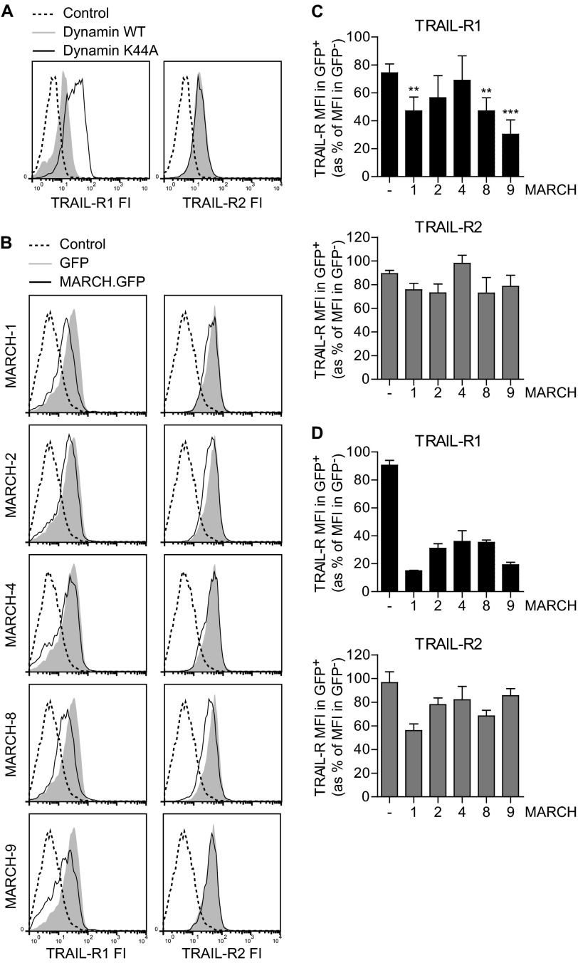
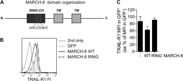
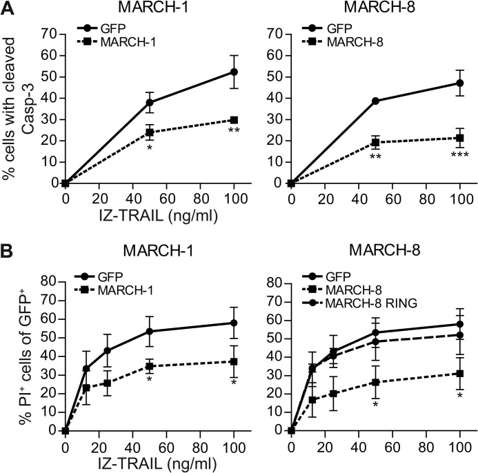
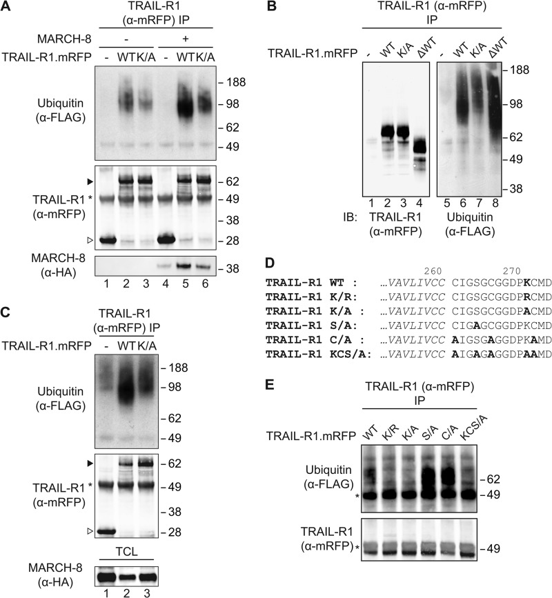
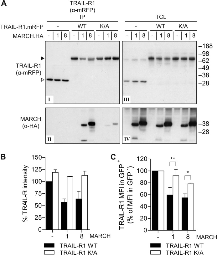
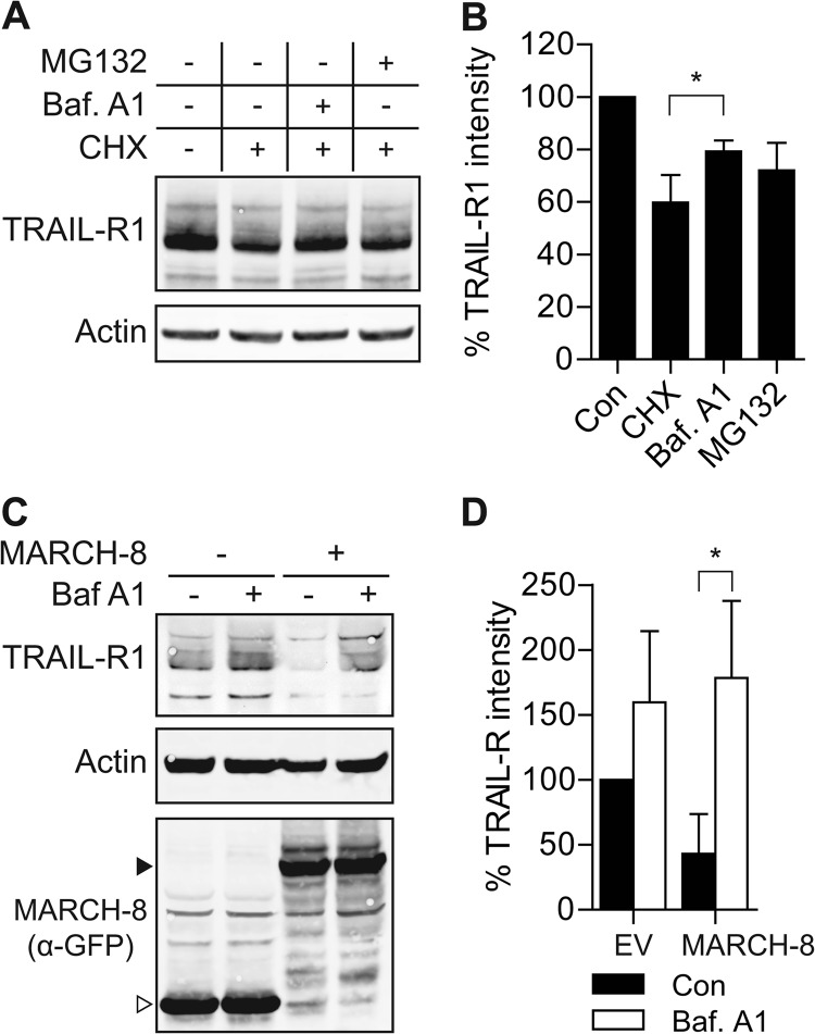
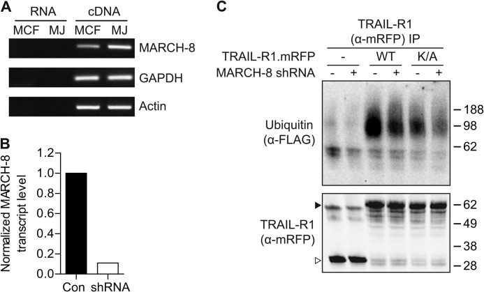
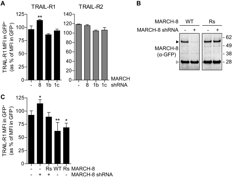
The eleven members of the membrane-associated RING-CH (MARCH) ubiquitin ligase family are relatively unexplored. Upon exogenous (over)expression, a number of these ligases can affect the trafficking of membrane molecules. However, only for MARCH-1 endogenous functions have been demonstrated. For the other endogenous MARCH proteins, no functions or substrates are known. We report here that TRAIL-R1 is a physiological substrate of the endogenous MARCH-8 ligase. Human TRAIL-R1 and R2 play a role in immunosurveillance and are targets for cancer therapy, because they selectively induce apoptosis in tumor cells. We demonstrate that TRAIL-R1 is down-regulated from the cell surface, with great preference over TRAIL-R2, by exogenous expression of MARCH ligases that are implicated in endosomal trafficking, such as MARCH-1 and -8. MARCH-8 attenuated TRAIL-R1 cell surface expression and apoptosis signaling by virtue of its ligase activity. This suggested that ubiquitination of TRAIL-R1 was instrumental in its down-regulation by MARCH-8. Indeed, in cells with endogenous MARCH expression, TRAIL-R1 was ubiquitinated at steady-state, with the conserved membrane-proximal lysine 273 as one of the potential acceptor sites. This residue was also essential for the interaction of TRAIL-R1 with MARCH-1 and MARCH-8 and its down-regulation by these ligases. Gene silencing identified MARCH-8 as the endogenous ligase that ubiquitinates TRAIL-R1 and attenuates its cell surface expression. These findings reveal that endogenous MARCH-8 regulates the steady-state cell surface expression of TRAIL-R1.


Figures

FIGURE 1.
FIGURE 1.
MARCH proteins preferentially down-regulate TRAIL-R1 cell surface levels in MCF-7Casp-3 cells. A, MCF-7Casp-3 breast cancer cells were transfected to express WT or K44A dynamin together with GFP. To detect endogenous TRAIL-R1 or TRAIL-R2 at the cell surface, the cells were stained with specific antibodies and a second step reagent, or with second step reagent only (Control) followed by flow cytometric analysis. Histograms of TRAIL-R fluorescence intensity (FI) in the GFP positive populations are shown. B and C, MCF-7Casp-3 cells were transfected to express GFP-tagged MARCH-1, -2, -4, -8, -9, or GFP only (Control) and cell surface levels of TRAIL-R1 (left) and TRAIL-R2 (right) were determined by flow cytometry. Representative histograms of TRAIL-R intensity in the GFP positive populations are shown in B. Panel C shows the quantification of TRAIL-R1 and -R2 expression in MCF-7Casp-3 cells expressing the indicated MARCH-GFP proteins or GFP only (−). The MFI, denoting TRAIL-R cell surface levels in GFP+ cells expressing MARCH-GFP or GFP only is expressed as percentage of the MFI in untransfected GFP cells in the same cell population. Data represent mean ± S.D. of values from at least 3 independent experiments. Asterisks indicate statistically significant differences between MARCH-transfected and GFP-transfected control cells (one-way analysis of variance, Bonferroni correction; *, p < 0.05; **, p < 0.01; ***, p < 0.001). D, this experiment was performed and quantified as outlined in B and C, but in this case using the melanoma cell line Mel JuSo. Data represent mean ± S.D. of values from 2 independent experiments.
FIGURE 2.
FIGURE 2.
MARCH-8 requires a functional RING domain to down-regulate TRAIL-R1 cell surface expression. MCF-7Casp-3 cells were transfected to express GFP only (−), or GFP-tagged WT MARCH-8, or a MARCH-8 variant carrying ligase-inactivating mutations in its RING domain (MARCH-8 RING). Cell surface levels of TRAIL-R1 were determined by antibody staining, followed by flow cytometric analysis. A, schematic depiction of MARCH-8, on a relative scale, with indication of the RING-CH domain and transmembrane (TM) segments, as well as the three point mutations. B, primary data from a representative experiment, showing histograms of TRAIL-R1 cell surface expression (fluorescence intensity, FI) in GFP+ (MARCH-transfected) cells. C, TRAIL-R1 cell surface expression was quantified and statistically analyzed as described in the legend to Fig. 1C. Data represent mean ± S.D. of values from 3 independent experiments. Asterisk indicates statistically significant differences between cells with WT MARCH-8 versus control or the RING mutant (one-way analysis of variance, Bonferroni correction; *, p < 0.05).
FIGURE 3.
FIGURE 3.
MARCH overexpression inhibits apoptosis induction by TRAIL. A, MCF-7Casp-3 cells were transfected to express either GFP only, or GFP-tagged MARCH-1, or -8. At 24 h after transfection, cells were stimulated with IZ-TRAIL for 5 h and caspase-3 cleavage was determined by flow cytometry in the GFP positive cell populations. B, as in A, with the following adaptations. Cells were transfected with WT MARCH-1 or -8, or with the MARCH-8 RING mutant described in the legend to Fig. 2. Cells were stimulated with TRAIL for 14 h, and cell death was read out by propidium iodide (PI) uptake. Data in A and B represent mean ± S.D. of values from 3 to 4 independent experiments. The percentage of cells with cleaved caspase-3 or PI uptake in the untreated control samples was subtracted. Asterisks indicate statistically significant differences between MARCH.GFP-transfected and GFP only-transfected control cells at the indicated concentration of TRAIL (Student's t test; *, p < 0.05; **, p < 0.01; ***, p < 0.001).
FIGURE 4.
FIGURE 4.
Steady-state ubiquitination of TRAIL-R1 on lysine residue 273 by an endogenous machinery. A, MCF-7Casp-3 cells were transfected to express FLAG-ubiquitin, together with mRFP only (−), or with mRFP-chimeras of WT TRAIL-R1 (WT) or its K273A lysine mutant (K/A). MARCH-8.HA cDNA (+) or an empty control vector (−) were additionally transfected as indicated. Cells were lysed in Nonidet P-40 buffer, TRAIL-R1 was isolated with anti(α)-mRFP antibody and immunoprecipitates (IP) were analyzed by immunoblotting (IB) with α-mRFP antibody to detect TRAIL-R1, α-FLAG antibody to detect ubiquitin and α-HA antibody to detect MARCH-8. Asterisk denotes the heavy chain of the antibody used for IP. Solid and open arrowheads indicate, respectively, TRAIL-R1.mRFP and mRFP only. Blot is representative of 4 independent experiments. B, MCF-7Casp-3 cells were transfected to express FLAG-ubiquitin, together with either mRFP only (−), with mRFP chimeras of WT TRAIL-R1 (WT) or the K273A TRAIL-R1 mutant (K/A), or with a truncated TRAIL-R1 lacking the C-terminal 116 residues (ΔWT). TRAIL-R1 was isolated with α-mRFP antibody and immunoprecipitates were analyzed by immunoblotting with α-mRFP antibody to detect TRAIL-R1 and with α-FLAG antibody to detect ubiquitin. Data shown are representative of two independent experiments. C, MCF-7Casp-3 cells were transfected to express FLAG-ubiquitin, together with mRFP only (−), or with mRFP-chimeras of WT TRAIL-R1 (WT) or its K273A lysine mutant (K/A). Cells were lysed by boiling in SDS, Nonidet P-40 buffer was added in excess and immunoprecipitation of TRAIL-R1 and analysis were performed as outlined for panel A. Asterisk denotes the heavy chain of the antibody used for IP. Solid and open arrowheads indicate, respectively, TRAIL-R1.mRFP and mRFP only. The blot is representative of 2 independent experiments. D, alignment of primary amino acid sequence of part of the transmembrane segment (italic) and the remaining 14 residues of the cytoplasmic tail of the truncated TRAIL-R1 mutants used in E. Relevant potential ubiquitination sites are shown in bold. E, MCF-7Casp-3 cells were transfected to express FLAG-ubiquitin, together with mRFP-tagged TRAIL-R1 WT or mutants shown in D. TRAIL-R1 was isolated with α-mRFP antibody and immunoprecipitates were analyzed by immunoblotting with α-mRFP antibody to detect TRAIL-R1 and α-FLAG antibody to detect ubiquitin. The blot is representative of 2 independent experiments. Asterisk denotes the heavy chain of the antibody used for IP.
FIGURE 5.
FIGURE 5.
MARCH-1 and -8 interact with and down-regulate wild-type TRAIL-R1, but not the TRAIL-R1 K273A mutant. A, MCF-7Casp-3 cells were transfected to express mRFP only (−), mRFP-tagged WT TRAIL-R1 or K273A (K/A) mutant, together with HA-tagged MARCH-1, MARCH-8, or empty vector (−), as indicated. Immunoprecipitation was performed with α-mRFP antibody and immunoprecipitates (IP) were analyzed by immunoblotting with α-mRFP and α-HA antibodies to detect TRAIL-R1 and MARCH-1/8, respectively. Panel I, mRFP detection in IP of TRAIL-R1.mRFP and control mRFP; panel II, MARCH-1 and -8 detection in IP of TRAIL-R1.mRFP and control mRFP; panel III, mRFP detection (TRAIL-R1.mRFP or RFP only) in total cell lysates (TCL); panel IV, MARCH-1 and -8 detection in TCL. B, quantification of TRAIL-R1 down-regulation in total cell lysates. Total protein levels of WT TRAIL-R1 and the K/A mutant in TCL of control cells (−), or those expressing HA-tagged MARCH-1 or -8 were quantified from Western blots as depicted in panel III of A and plotted as percentage of the WT TRAIL-R1.mRFP expression in control cells. Data represent mean ± S.D. of values from the experiment depicted in A and 2 additional experiments. C, impact of MARCH-1 or MARCH-8 on WT and K/A mutant TRAIL-R1 cell surface expression. Cells stably expressing WT or K273A TRAIL-R1.mRFP were transfected to express GFP (−), GFP-tagged MARCH-1 or MARCH-8, and stained with antibody to TRAIL-R1 as outlined for Fig. 2. Quantification of 2–4 independent experiments assessing TRAIL-R1 MFI in GFP+ cells as the percentage of TRAIL-R1 MFI in GFP cells, whereby the values in control cells were set at 100%. Values represent mean ± S.D. Asterisks indicate statistically significant differences between TRAIL-R1 WT or K/A mutant (Student's t test; *, p < 0.05, **, p < 0.01).
FIGURE 6.
FIGURE 6.
MARCH-8 targets endogenous TRAIL-R1 for lysosomal degradation. A and B, H358 cells were treated with cycloheximide (CHX; 50 μg/ml) alone, or with CHX in combination with bafilomycin A1 (Baf A1; 200 nm) or MG132 (10 μm) for 16 h. Total cell lysates were analyzed by immunoblotting with α-TRAIL-R1 and α-actin antibodies. A, data of a representative experiment. B, mean ± S.D. of quantified values from 3 independent experiments. TRAIL-R1 intensity was corrected for actin expression, the control value was set to 100%. Asterisk indicates statistically significant difference (Student's t test; *, p < 0.05). C and D, MCF-7Casp-3 cells were transfected to express GFP only (−), or GFP-tagged MARCH-8. Cells were treated with BafA1 (100 nm) or left untreated for 16 h. Total cell lysates of GFP+ cells, obtained by flow cytometric sorting, were analyzed by immunoblotting with α-TRAIL-R1 and α-actin antibodies, and α-GFP antibody to detect MARCH-8. Solid and open arrowheads indicate, respectively, MARCH-8.GFP and GFP only. C, data of a representative experiment. D, mean ± S.D. of quantified values from 3 independent experiments. TRAIL-R1 intensity was corrected for actin expression, control value was set to 100%. Asterisk indicates a statistically significant difference (Student's t test; *, p < 0.05).
FIGURE 7.
FIGURE 7.
TRAIL-R1 is a substrate of endogenous MARCH-8. A, endogenous MARCH-8 expression in MCF-7Casp-3 (MCF-7) and Mel Juso (MJ) cells, as determined by RT-PCR on cDNA. Non-reverse transcribed RNA (RNA) was used as a control template to exclude amplification of genomic DNA. B, down-regulation of endogenous MARCH-8 by RNAi. MCF-7Casp-3 cells were transfected with MARCH-8 shRNA, together with GFP. MARCH-8 shRNA expressing cells (GFP+) and nonexpressing cells (GFP) cells were separated by flow cytometric sorting and analyzed for endogenous MARCH-8 transcript levels by quantitative RT-PCR. Signals that were corrected for GAPDH and MARCH-8 transcript levels in the GFP+ population were normalized to the levels in the GFP population. C, MCF-7Casp-3 cells were transfected to express mRFP alone, WT TRAIL-R1.mRFP, or K273A TRAIL-R1.mRFP, together with FLAG-ubiquitin and either an empty vector, or the MARCH-8 targeting shRNA. TRAIL-R1 was isolated by immunoprecipitation with α-mRFP antibody and immunoprecipitates (IP) were analyzed by immunoblotting for TRAIL-R1 (α-mRFP) or ubiquitin (α-FLAG). Solid and open arrowheads indicate, respectively, TRAIL-R1.mRFP and mRFP only. Data shown are representative of 3 independent experiments.
FIGURE 8.
FIGURE 8.
Endogenous MARCH-8 regulates cell surface expression of endogenous TRAIL-R1. A, MCF-7Casp-3 cells were transfected to express GFP (−), together with either a control vector (−), with MARCH-8 targeting shRNA (8), or with two different MARCH-1-targeting shRNAs (1b and 1c). Endogenous TRAIL-R1 cell surface expression was determined by flow cytometric analysis and data were evaluated as described in the legend to Fig. 2. Data represent mean values ± S.D. from 3 independent experiments. Asterisks indicate statistically significant differences compared with GFP-transfected control cells (one-way analysis of variance, Bonferroni correction; ***, p < 0.001). B, validation of the MARCH-8 rescue construct. MCF-7Casp-3 cells were transfected to express GFP-tagged MARCH-8 WT, or a MARCH-8 rescue (Rs) variant carrying silent mutations to allow escape from RNAi. Cells were cotransfected with empty vector (−) or MARCH-8 shRNA construct. MARCH-8.GFP expression was analyzed by immunoblotting for GFP in total cell lysates. C, MCF-7Casp-3 cells were transfected to express GFP (−), GFP-tagged MARCH-8 WT or Rs alone (−), or in conjunction MARCH-8 targeting shRNA (+). Endogenous TRAIL-R1 cell surface expression was determined by flow cytometric analysis and data were evaluated as described in the legend to Fig. 2. Data represent mean ± S.D. from at least 4 independent experiments. Asterisks indicate statistically significant differences compared with GFP-transfected control cells (one-way analysis of variance, Bonferroni correction; ***, p < 0.001).

Similar articles

Cited by

References

    1. Smyth M. J., Cretney E., Takeda K., Wiltrout R. H., Sedger L. M., Kayagaki N., Yagita H., Okumura K. (2001) Tumor necrosis factor-related apoptosis-inducing ligand (TRAIL) contributes to interferon γ-dependent natural killer cell protection from tumor metastasis. J. Exp. Med. 193, 661–670 - PMC - PubMed
    1. Takeda K., Hayakawa Y., Smyth M. J., Kayagaki N., Yamaguchi N., Kakuta S., Iwakura Y., Yagita H., Okumura K. (2001) Involvement of tumor necrosis factor-related apoptosis-inducing ligand in surveillance of tumor metastasis by liver natural killer cells. Nat. Med. 7, 94–100 - PubMed
    1. Grosse-Wilde A., Voloshanenko O., Bailey S. L., Longton G. M., Schaefer U., Csernok A. I., Schütz G., Greiner E. F., Kemp C. J., Walczak H. (2008) TRAIL-R deficiency in mice enhances lymph node metastasis without affecting primary tumor development. J. Clin. Invest. 118, 100–110 - PMC - PubMed
    1. Ashkenazi A., Herbst R. S. (2008) To kill a tumor cell. The potential of proapoptotic receptor agonists. J. Clin. Invest. 118, 1979–1990 - PMC - PubMed
    1. Kimberley F. C., Screaton G. R. (2004) Following a TRAIL. Update on a ligand and its five receptors. Cell Res. 14, 359–372 - PubMed

Publication types

MeSH terms

Substances

LinkOut - more resources