. 2013 Feb 15;288(7):5081-9.
doi: 10.1074/jbc.M112.447029. Epub 2013 Jan 7.

Drosophila heparan sulfate 6-O-endosulfatase Sulf1 facilitates wingless (Wg) protein degradation

Affiliations

Drosophila heparan sulfate 6-O-endosulfatase Sulf1 facilitates wingless (Wg) protein degradation

Adam Kleinschmit et al. J Biol Chem. .

Abstract

Heparan sulfate proteoglycans regulate various physiological and developmental processes through interactions with a number of protein ligands. Heparan sulfate (HS)-ligand binding depends on the amount and patterns of sulfate groups on HS, which are controlled by various HS sulfotransferases in the Golgi apparatus as well as extracellular 6-O-endosulfatases called "Sulfs." Sulfs are a family of secreted molecules that specifically remove 6-O-sulfate groups within the highly sulfated regions on HS. Vertebrate Sulfs promote Wnt signaling, whereas the only Drosophila homologue of Sulfs, Sulf1, negatively regulates Wingless (Wg) signaling. To understand the molecular mechanism for the negative regulation of Wg signaling by Sulf1, we studied the effects of Sulf1 on HS-Wg interaction and Wg stability. Sulf1 overexpression strongly inhibited the binding of Wg to Dally, a potential target heparan sulfate proteoglycan of Sulf1. This effect of Drosophila Sulf1 on the HS-Wg interaction is similar to that of vertebrate Sulfs. Using in vitro, in vivo, and ex vivo systems, we show that Sulf1 reduces extracellular Wg protein levels, at least partly by facilitating Wg degradation. In addition, expression of human Sulf1 in the Drosophila wing disc lowers the levels of extracellular Wg protein, as observed for Drosophila Sulf1. Our study demonstrates that vertebrate and Drosophila Sulfs have an intrinsically similar activity and that the function of Sulfs in the fate of Wnt/Wg ligands is context-dependent.


Figures

FIGURE 1.
FIGURE 1.
Sulf1 reduces Wg binding to Dally in vitro. A, effect of Sulf1 on Wg-Dally interaction. S2R+ or S2R+-pAW-Sulf-HA cell lines were transiently transfected with empty vector, sec-dally-myc, and/or wg cDNA. After 3 days, the cultured medium was collected and incubated with anti-Myc-conjugated protein G-agarose. After a 24-h incubation, Sec-Dally-Myc was immunoprecipitated, and Wg levels in the precipitates were analyzed by immunoblotting (IB) using anti-Wg antibody. Conditioned medium samples taken before the incubation with anti-Myc-agarose were loaded on the left lanes (Input). Note that Wg protein levels are substantially reduced in the Dally immunoprecipitates (IP) derived from the Sulf1-expressing cells. B, Wg-Dally binding assay in Sulf1-inducible cells. S2R+-pMt-Sulf1-HA cells were transiently transfected with empty vector, sec-dally-myc, and/or wg cDNA. The same immunoprecipitation methodology as above was used to detect Wg-Dally binding.
FIGURE 2.
FIGURE 2.
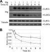
Sulf1 reduces Wg levels in cultured cells. A, the levels of Wg monitored over time in S2R+-pMt-Sulf1-HA cell line. Wg protein from Wg conditioned medium was allowed to bind to the cells at 4 °C for 1 h. After washing away unbound Wg and incubating the cells with or without CuSO4 at 25 °C, the remaining Wg protein was detected from cell lysates at each respective time point above, with α-tubulin as a loading control. B, graph representing the averaged densitometry values (t = 0 set to 1) from four independent experiments as described above. Arbitrary optical densitometry unit values for non-induced (solid line) and induced (dotted line) cells were calculated using ImageJ and normalized to α-tubulin, which was used as a loading control. Error bars for each averaged time point were calculated using standard error (S.D./square root (n), n = 4).
FIGURE 3.
FIGURE 3.
Cellular degradation contributes to Sulf1-dependent decrease in the level of Wg. A, effects of bafilomycin treatment on Wg protein levels. In vitro Wg protein assay was performed as in Fig. 2 using S2R+-pMt-Sulf1-HA cells with (Sulf1 +) or without (Sulf1 −) induction of Sulf1 expression. The cells were allowed to bind Wg, and unbound Wg was washed away with medium. After incubation for the indicated times, Wg protein was detected from cell lysates. The same experiment was performed in the absence (top) or the presence (bottom) of 25 nm bafilomycin. B, effects of rab 7 RNAi treatment on Wg protein levels. S2R+-pMt-Sulf1-HA cells with (Sulf1 +) or without (Sulf1 −) induction of Sulf1 expression were transfected with double-stranded RNA (dsRNA) for rab7 (bottom). Cells with no dsRNA treatment were used as a control (top). After incubation for the indicated times, Wg protein levels were monitored by immunoblotting.
FIGURE 4.
FIGURE 4.
Wg in the conditioned medium. Lanes 1–3, a dilution series of soluble Wg protein in the conditioned medium of wg-expressing cells (S2-tub-Wg) was precipitated with TCA. The amount of Wg identical to that on the cell surface at time 0 was determined as 100%. The amount of Wg loaded in lanes 1, 2, and 3 corresponds to 10, 5, and 2.5%, respectively. Lanes 4–7, control (lanes 4 and 5) and Sulf1-expressing (lanes 6 and 7) cells were incubated with Wg, and unbound Wg was washed away. At time 0 (lanes 4 and 6) and after a 2-h induction (lanes 5 and 7), proteins in the conditioned medium (supernatant, sup) were precipitated by TCA, and Wg in the precipitates was detected by immunoblotting.
FIGURE 5.
FIGURE 5.
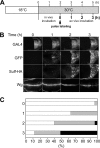
Induction of Sulf1 reduces extracellular Wg gradient. A, graphic depicting the GAL4/GAL80ts system. A temperature-sensitive transgenic allele of GAL80 (black oval) driven ubiquitously by the tubulin promotor (tub-GAL80ts) actively binds to GAL4 (gray oval) in a dimer-dimer interaction at the permissive temperature (18 °C), repressing target gene expression (Sulf1-HA). After shifting to the restrictive temperature (30 °C), GAL80ts loses its affinity to the GAL4 activation domain, and target gene expression is turned on. B, immunostaining of mid-third instar larval wing discs showing the gradual induction (2–8.5 h) of Sulf1 after shifting whole larvae to the GAL80ts restrictive temperature. Expression of GAL4 protein and GFP at each time point is also shown. GAL4 protein was detected at a constant level throughout the time course. C, graphic depiction of Wg gradient phenotype penetrance. Bar graphs show the percentage of wing discs exhibiting wild-type (white), mild (gray), and strong (black) phenotypes at each respective time point after transferring whole larvae to the restrictive temperature. Examples of each phenotypic category are shown on the left. Signal intensity of three discs is shown in pseudocolor. Pseudocolor scale ranges from white (highest signal intensity) to dark blue (lowest signal intensity). The number of discs classified into each phenotypic category at respective time points is shown in Table 1. A, anterior; P, posterior.
FIGURE 6.
FIGURE 6.
Pulse-chase analysis of extracellular Wg using ex vivo culture system. A, graphic depicting Wg ex vivo pulse-chase assay system. tub-GAL80ts/+; hh-GAL4 UAS-GFP/UAS-Sulf1-HA larvae were reared at 18 °C (GAL80ts permissive temperature). At mid-third instar larval stage, the culture was transferred to 30 °C (GAL80ts restrictive temperature) and further incubated in vivo. After 2 h, wing discs were dissected, and extracellular Wg was pulse-labeled with anti-Wg antibody. Unbound antibody was cleared by washing, and tissue was incubated ex vivo at 30 °C for 0–3 h. B, confocal images of ex vivo cultured discs stained for GAL4, GFP, Sulf1-HA, and extracellular Wg. Discs cultured ex vivo for 0–3 h after pulse labeling are shown. GAL4 was constitutively expressed throughout the assay, whereas expression levels of GFP, marking the GAL4-active domain, and Sulf1-HA increased over time. An example of extracellular Wg staining is shown for each time point. The extracellular Wg gradient exhibited a predominantly wild-type phenotype in the posterior compartment at 0 and 1 h, whereas the majority of sample discs exhibited a mild phenotype and a strong phenotype at 2 and 3 h, respectively. C, graphical depiction of the extracellular Wg phenotype observed at 0–3-h time points of the ex vivo Wg pulse-chase assay. Bar graphs show the percentage of wing discs exhibiting wild-type (white), mild (gray), and strong (black) phenotypes at each respective time point. The number of discs classified into each phenotypic category at respective time points is shown in Table 2.
FIGURE 7.
FIGURE 7.
Human Sulf1 reduces extracellular Wg in the Drosophila wing disc. A, A′, B, and B′, extracellular Wg staining (magenta) of wing discs overexpressing Drosophila (A and A′) and human (B and B′) Sulf1. Expression of UAS-Sulf1 and UAS-hSulf1 was induced in the posterior compartment by an hh-Gal4 driver. The posterior compartment is marked by GFP expression (A′ and B′, arrows in A and B). C, C′, D, and D′, immunostaining of extracellular Wg (magenta) in wing discs bearing FLP-OUT clones overexpressing Drosophila (C and C′) and human (D and D′) Sulf1. Extracellular levels of Wg protein are reduced in the FLP-OUT clones (arrows) marked with GFP (B′ and C′).
FIGURE 8.
FIGURE 8.
A model for the regulation of Wnt/Wg signaling by vertebrate and Drosophila Sulf1. In vertebrates, Wnt ligands (red) show high affinity to a binding site on HS, which presumably includes a 6-O-sulfate group. 6-O-desulfation by Sulf1 (blue) converts HS to a low affinity binding state, which can present Wnt to receptor (R) (magenta) (10). In Drosophila, Sulf1 activity releases Wg from cell surface HS similarly to vertebrate systems. However, a major fraction of Wg dissociated from HS is more quickly internalized and degraded.

Similar articles

Cited by

References

    1. Esko J. D., Selleck S. B. (2002) Order out of chaos: assembly of ligand binding sites in heparan sulfate. Annu. Rev. Biochem. 71, 435–471 - PubMed
    1. Kirkpatrick C. A., Selleck S. B. (2007) Heparan sulfate proteoglycans at a glance. J. Cell Sci. 120, 1829–1832 - PubMed
    1. Tabata T., Takei Y. (2004) Morphogens, their identification and regulation. Development 131, 703–712 - PubMed
    1. Kirkpatrick C. A., Knox S. M., Staatz W. D., Fox B., Lercher D. M., Selleck S. B. (2006) The function of a Drosophila glypican does not depend entirely on heparan sulfate modification. Dev. Biol. 300, 570–582 - PubMed
    1. Bernfield M., Götte M., Park P. W., Reizes O., Fitzgerald M. L., Lincecum J., Zako M. (1999) Functions of cell surface heparan sulfate proteoglycans. Annu. Rev. Biochem. 68, 729–777 - PubMed

Publication types

LinkOut - more resources