. 2013 Mar 1;288(9):6438-50.
doi: 10.1074/jbc.M112.412478. Epub 2013 Jan 8.

Post-synaptic density-95 (PSD-95) binding capacity of G-protein-coupled receptor 30 (GPR30), an estrogen receptor that can be identified in hippocampal dendritic spines

Affiliations

Post-synaptic density-95 (PSD-95) binding capacity of G-protein-coupled receptor 30 (GPR30), an estrogen receptor that can be identified in hippocampal dendritic spines

Keith T Akama et al. J Biol Chem. .

Abstract

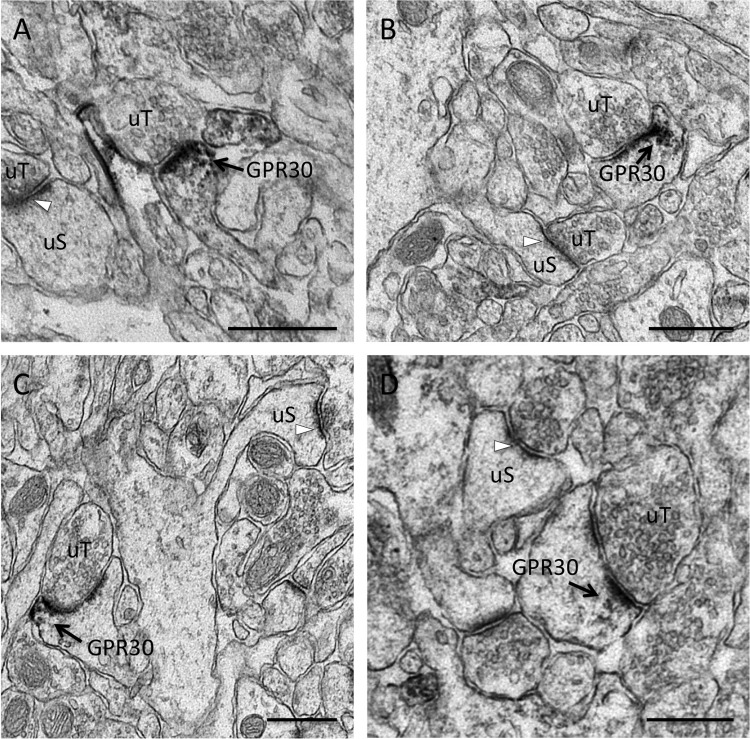
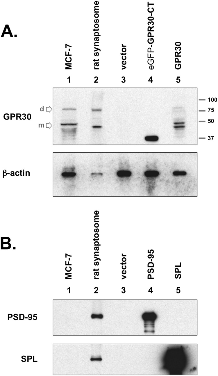
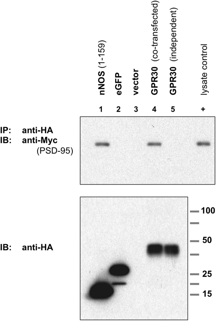
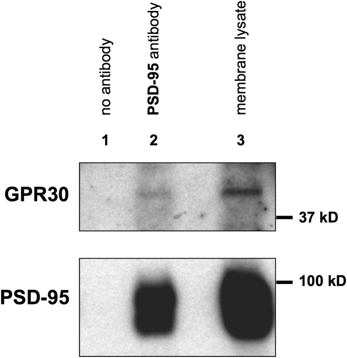
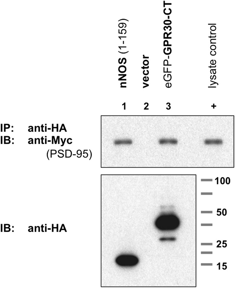
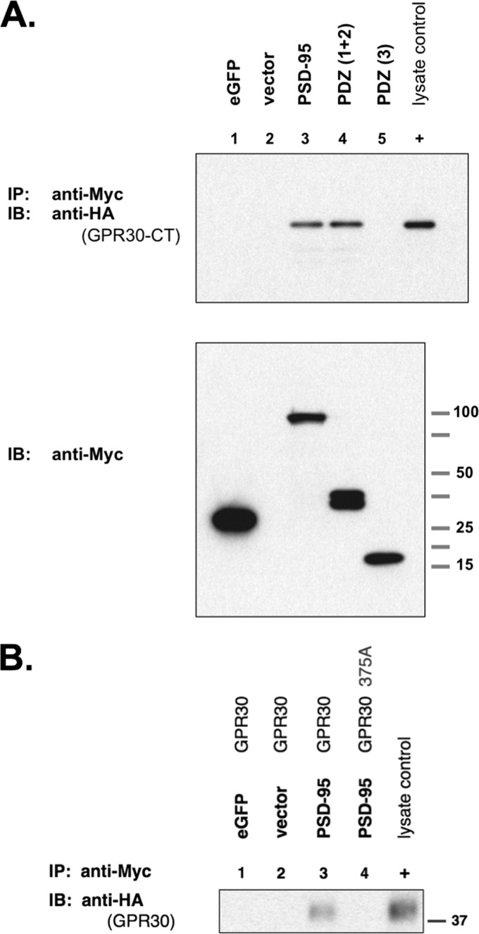
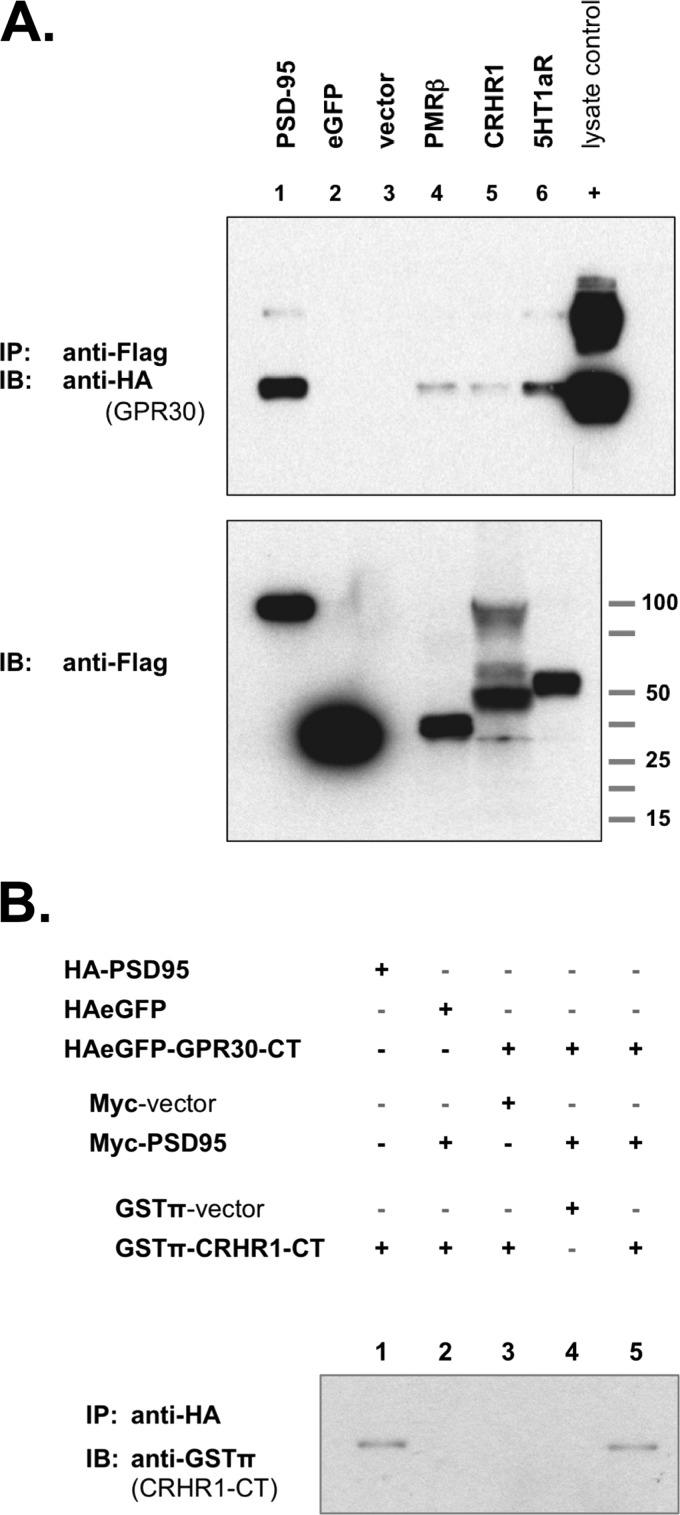
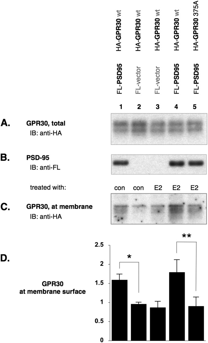
The estrogen 17β-estradiol (E2) modulates dendritic spine plasticity in the cornu ammonis 1 (CA1) region of the hippocampus, and GPR30 (G-protein coupled estrogen receptor 1 (GPER1)) is an estrogen-sensitive G-protein-coupled receptor (GPCR) that is expressed in the mammalian brain and in specific subregions that are responsive to E2, including the hippocampus. The subcellular localization of hippocampal GPR30, however, remains unclear. Here, we demonstrate that GPR30 immunoreactivity is detected in dendritic spines of rat CA1 hippocampal neurons in vivo and that GPR30 protein can be found in rat brain synaptosomes. GPR30 immunoreactivity is identified at the post-synaptic density (PSD) and in the adjacent peri-synaptic zone, and GPR30 can associate with the spine scaffolding protein PSD-95 both in vitro and in vivo. This PSD-95 binding capacity of GPR30 is specific and determined by the receptor C-terminal tail that is both necessary and sufficient for PSD-95 interaction. The interaction with PSD-95 functions to increase GPR30 protein levels residing at the plasma membrane surface. GPR30 associates with the N-terminal tandem pair of PDZ domains in PSD-95, suggesting that PSD-95 may be involved in clustering GPR30 with other receptors in the hippocampus. We demonstrate that GPR30 has the potential to associate with additional post-synaptic GPCRs, including the membrane progestin receptor, the corticotropin releasing hormone receptor, and the 5HT1a serotonin receptor. These data demonstrate that GPR30 is well positioned in the dendritic spine compartment to integrate E2 sensitivity directly onto multiple inputs on synaptic activity and might begin to provide a molecular explanation as to how E2 modulates dendritic spine plasticity.


Figures

FIGURE 1.
FIGURE 1.
GPR30 immunoreactivity in CA1 dendritic spine profiles. Shown are representative electron micrographs of GPR30 immunolabeling from the adult female rat CA1 hippocampus. Using pre-embedding electron microscopy, GPR30-labeled dendritic spines (GPR30) are identified by diffuse granular immuno-peroxidase reaction product and are indicated by black arrows. Unlabeled spines (uS) with unlabeled PSDs (white arrows) and unlabeled pre-synaptic terminals (uT) also are indicated. GPR30 IR is evident in multiple spine profile types, including thin spines (A), mushroom-shaped spines (B), cupped spines (C), and spines with perforated PSDs (D). GPR30-IR localized both at the PSD as well as in the peri-synaptic zone of the PSD. Panel A, at proestrus. Panels B–D, at diestrus. Bars, 500 nm.
FIGURE 2.
FIGURE 2.
GPR30 protein expression in the rat brain. A, upper blot, GPR30 was detected in crude rat brain synaptosome preparations (lane 2). COS-7 cells did not express GPR30, as GPR30 was not detected in empty vector-transfected COS-7 whole cell lysates (lane 3). GPR30 (C-terminal-specific) antibody recognizes the C-terminal domain of GPR30 (GPR30-CT), which was subcloned for chimeric expression with eGFP (lane 4). The rat full-length GPR30 protein are also detected when exogenously expressed in COS-7 cells (lane 5). MCF-7 whole cell lysate was used as a positive GPR30 antibody control (lane 1). The primary lower molecular weight GPR30 band is the monomeric protein state (m), and the upper secondary band at approximately twice the apparent molecular weight is likely the dimeric state (d) for the receptor. The apparent molecular weight ladder is shown on right. Lower blot, the same lysates were immunoblotted (IB) for actin protein content. Both blots are representative of five independent experiments. B, rat brain preparations contain synaptosomal proteins, including the PSD proteins PSD-95 and SPL. MCF-7 whole cell lysate (lane 1), rat brain synaptosome lysate (lane 2), or COS-7 whole cell lysates (lane 3, empty vector; lane 4, Myc-PSD95 transfected; lane 5, Myc-SPL transfected) were immunoblotted for PSD-95 protein (upper blot) or for SPL protein (lower blot). Blots are representative of three independent experiments.
FIGURE 3.
FIGURE 3.
GPR30 associates with PSD-95 and requires co-expression. Upper blot, PSD-95 co-IP with GPR30 is shown. COS-7 cells were transiently co-transfected to express Myc-PSD95 plus the lane-indicated HA-tagged proteins. The N-terminal 159 amino acids (1–159) of nNOS, HA-nNOS-(1–159) (lane 1), HA-eGFP (lane 2), empty HA vector (lane 3), and HA-tagged murine GPR30 (lane 4) are shown. Additionally, cells were transfected independently with HA-GPR30 and Myc-PSD95, and whole cell lysates from both transfections were combined before immunoprecipitation. HA-tagged proteins were immunoprecipitated with an anti-HA antibody, and PSD-95 protein was detected by immunoblot (IB) with an anti-Myc antibody. HA-nNOS-(1–159) served as a PSD-95 co-IP-positive control, and HA-eGFP served as a PSD-95 co-IP negative control. When co-expressed in the same cell, GRP30 was able to associate with and to co-immunoprecipitate PSD-95 (lane 4), but when expressed independently, GPR30 was unable to co-immunoprecipitate PSD-95 (lane 5). 0.1% of whole cell lysate from Myc-PSD95-transfected COS-7 cells was also run as a positive Myc-epitope control (+). Lower blot, HA-tagged proteins in whole cell lysates. Blots are representative of five independent experiments.
FIGURE 4.
FIGURE 4.
GPR30 receptor co-immunoprecipitates in vivo with PSD-95 protein from membrane preparations isolated from the adult rat hippocampus. Whole hippocampi were dissected from adult female rats, and hippocampal membrane fractions were isolated by sucrose density gradients and high speed ultracentrifugation. Membrane lysates were incubated with either anti-PSD-95 antibody (lane 2) or with mouse IgG only (lane 1, no antibody). Immunoprecipitates were then immunoblotted with anti-GPR30 antibody (upper blot) or with anti-PSD-95 antibody (lower blot). Likewise, whole membrane lysate (1% of total protein amount used in the immunoprecipitation step in the upper blot, 0.1% total protein amount in the lower blot) was run alongside each immunoprecipitation (lane 3). GPR30 receptor co-immunoprecipitated with PSD-95 (lane 2) but not with IgG alone (lane 1). Blots are representative of three independent experiments.
FIGURE 5.
FIGURE 5.
The C-terminal tail of GPR30 is sufficient for PSD-95 association. Upper blot, PSD-95 co-immunoprecipitated with the GPR30 C-terminal tail (CT). Cells were co-transfected with Myc-PSD95 plus HA-tagged nNOS-(1–159) (lane 1), empty HA vector (lane 2), or HA-tagged eGFP-GPR30-CT, where the GPR30-CT is chimerically expressed with HA-eGFP (eGFP-GPR30-CT, lane 3). Anti-HA IP followed by anti-Myc immunoblot (IB) demonstrates that the GPR30-CT is sufficient for PSD-95 co-IP. 0.1% of whole cell lysate from Myc-PSD95-transfected COS-7 cells was also run as a positive Myc-epitope control (+). Lower blot, HA-tagged proteins in whole cell lysates are shown. Blots are representative of five independent experiments.
FIGURE 6.
FIGURE 6.
The C-terminal tail of GPR30 is necessary for PSD-95 association. The terminal carboxylate residue in GPR30 is valine at position 375. In both the full-length GPR30 protein as well as the GPR30-CT protein, this terminal residue was mutated into an alanine residue (375A) to disrupt the PDZ domain ligand sequence. Upper blot, COS-7 cells were co-transfected for Myc-PSD95 expression plus the indicated HA-tagged proteins: lane 1, full-length wild-type GPR30; lane 2, full-length GPR30 (375A); lane 3, eGFP-GPR30-CT (wt); lane 4, eGFP-GPR30 (375A). GPR30 (wt) and eGFP-GPR30-CT(wt) were able to co-immunoprecipitate PSD-95 protein (lanes 1 and 3), but GPR30 (375A) and eGFP-GPR30-CT (375A) with disrupted PDZ ligand sequences were unable to co-immunoprecipitate PSD-95 protein (lanes 2 and 4). 0.1% of whole cell lysate from Myc-PSD95-transfected COS-7 cells was also run as a positive Myc-epitope control (+). Lower blot, HA-tagged proteins in whole cell lysates are shown. Blots are representative of five independent experiments. IB, immunoblot.
FIGURE 7.
FIGURE 7.
PSD-95 associates with GPR30 via the N-terminal tandem PDZ domains. A, upper blot, GPR30 co-IP with PSD-95 is shown. COS-7 cells were co-transfected with HA-tagged eGFP-GPR30-CT plus the following Myc-tagged proteins: lane 1, eGFP; lane 2, empty Myc vector; lane 3, PSD-95; lane 4, the first two tandem PDZ domains of PSD-95, PDZ(1 + 2); lane 5, the third PDZ domain of PSD-95, PDZ(3). Whole cell lysates were collected, and Myc-tagged proteins were immunoprecipitated with an anti-Myc antibody. HA-tagged eGFP-GPR30-CT was immunoblotted with an anti-HA antibody. PSD-95 and PDZ(1 + 2) were able to co-immunoprecipitate the GPR30-CT (lanes 3 and 4), but the third PDZ(3) domain did not associate with GPR30-CT (lane 5). 0.1% of whole cell lysate from HA-eGFP-GPR30-CT-transfected COS-7 cells was also run as a positive HA-epitope control (+). Lower blot, Myc-tagged proteins in whole cell lysates are shown. Blots are representative of five independent experiments. B, full-length GPR30 can co-immunoprecipitate with PSD-95. COS-7 cells were co-transfected as indicated (lanes 1–4) with either Myc-tagged eGFP, Myc-vector, or Myc-tagged PSD-95 plus either HA-tagged full-length GPR30 or HA-tagged full-length GPR30375A mutant. Additionally, whole cell lysates prepared from HA-GPR30-transfected COS-7 cells were used as a positive lysate control (+) for immunoblotting. Myc-tagged proteins were immunoprecipitated from lysates with agarose-conjugated anti-Myc antibody, and the precipitates were then immunoblotted with a peroxidase-conjugated anti-HA antibody. Full-length GPR30 was able to co-immunoprecipitate with PSD-95 (lane 3), whereas the mutant full-length GPR30375A did not (lane 4). IB, immunoblot.
FIGURE 8.
FIGURE 8.
GPR30 associates with other GPCRs that have been previously identified in post-synaptic structures. A, upper blot, GPCR co-immunoprecipitates with GPR30. COS-7 cells were co-transfected with HA-tagged GPR30 plus the following FLAG-tagged expression constructs: lane 1, PSD-95; lane 2, eGFP; lane 3, empty FLAG vector; lane 4, PMRβ; lane 5, CRHR1, lane 6, 5HT1aR. FLAG-tagged proteins were precipitated with anti-FLAG antibody, and GPR30 protein was detected by immunoblot with anti-HA antibody. All three GPCRs could co-immunoprecipitate with GPR30 protein. 0.1% of whole cell lysate from HA-GPR30-transfected COS-7 cells was also run as a positive HA-epitope control (+). Lower blot, FLAG-tagged proteins in whole cell lysates are shown. Blots are representative of seven independent experiments. B, PSD-95 coupled GPR30-CT with CRHR1-CT. The C-terminal tail of the rat CRHR1 receptor was chimerically linked to GSTπ in a GSTπ-CRHR1-CT expression construct. COS-7 cells were transfected as indicated, and lysates were collected for precipitating HA-tagged proteins. When GPR30-CT was co-expressed with PSD-95 and with CRHR1-CT, GPR30-CT co-immunoprecipitated with CRHR1-CT (lane 5). In the absence of either GPR30-CT (lane 2) or PSD-95 (lane 3), this co-IP capability is lost. GPR30-CT coupling to CRHR1-CT is specific, as GPR30-CT and PSD-95 together cannot co-immunoprecipitate GSTπ alone (lane 4). As a control, PSD-95 alone can co-immunoprecipitate CRHR1-CT (lane 1). IB, immunoblot.
FIGURE 9.
FIGURE 9.
PSD-95 increases the amount of GPR30 residing at the plasma membrane. COS-7 cells were co-transfected and treated as indicated (A and C, diluent control (con), estrogen agonist (E2)). Surface proteins were biotinylated and affinity-precipitated with agarose-streptavidin. A, shown is an immunoblot (IB) on HA-tagged proteins before streptavidin affinity precipitation, therefore detecting the total amounts of GPR30 (biotinylated surface and non-biotinylated intracellular) per lysate. B, shown is an immunoblot on FLAG (FL-)tagged PSD-95 co-expression with HA-tagged GPR30. C, shown is an immunoblot for GPR30 on streptavidin affinity precipitation surface proteins. PSD-95 co-expression increased the amount of HA-tagged receptor available for cell surface biotinylation (lane 1 versus, lane 2, control-treated). In the absence of PSD-95 (lane 2 versus 3), agonist treatment had no effect to the level of GPR30 at the plasma membrane. Likewise, agonist treatment had no effect to the elevated level of GPR30 at the plasma membrane when PSD-95 was co-expressed (lanes 1 versus 4). When the C terminus was mutated (375A) and the ability of GPR30 to associate with PSD-95 was abolished, PSD-95 co-expression has no effect on the membrane levels of GPR30. D, shown is a graphics representation of panel C where GPR30 protein levels at the plasma membrane surface are expressed relative to untreated COS-7 cells co-transfected with GPR30 and PSD-95 (not shown). n = 3 independent experiments, ±S.D. *, statistically significant, p < 0.03. **, statistically significant, p < 0.02.

References

    1. McEwen B. S. (2001) Invited review. Estrogens effects on the brain. Multiple sites and molecular mechanisms. J. Appl. Physiol. 91, 2785–2801 - PubMed
    1. Srivastava D. P., Woolfrey K. M., Woolfrey K., Jones K. A., Shum C. Y., Lash L. L., Swanson G. T., Penzes P. (2008) Rapid enhancement of two-step wiring plasticity by estrogen and NMDA receptor activity. Proc. Natl. Acad. Sci. U.S.A. 105, 14650–14655 - PMC - PubMed
    1. Thomas P., Pang Y., Filardo E. J., Dong J. (2005) Identity of an estrogen membrane receptor coupled to a G protein in human breast cancer cells. Endocrinology 146, 624–632 - PubMed
    1. Carmeci C., Thompson D. A., Ring H. Z., Francke U., Weigel R. J. (1997) Identification of a gene (GPR30) with homology to the G-protein-coupled receptor superfamily associated with estrogen receptor expression in breast cancer. Genomics 45, 607–617 - PubMed
    1. Prossnitz E. R., Barton M. (2011) The G-protein-coupled estrogen receptor GPER in health and disease. Nat. Rev. Endocrinol 7, 715–726 - PMC - PubMed

Publication types