. 2013 Feb 22;288(8):5278-90.
doi: 10.1074/jbc.M112.409482. Epub 2013 Jan 8.

Interplay between calmodulin and phosphatidylinositol 4,5-bisphosphate in Ca2+-induced inactivation of transient receptor potential vanilloid 6 channels

Affiliations

Interplay between calmodulin and phosphatidylinositol 4,5-bisphosphate in Ca2+-induced inactivation of transient receptor potential vanilloid 6 channels

Chike Cao et al. J Biol Chem. .

Abstract

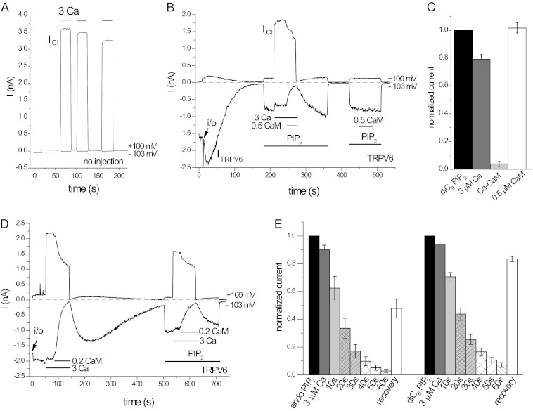
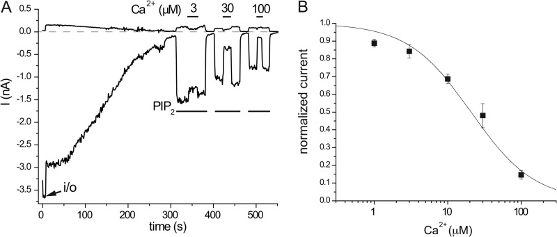
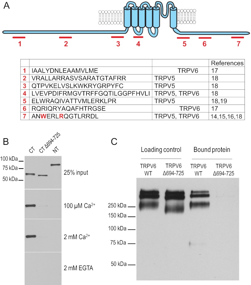
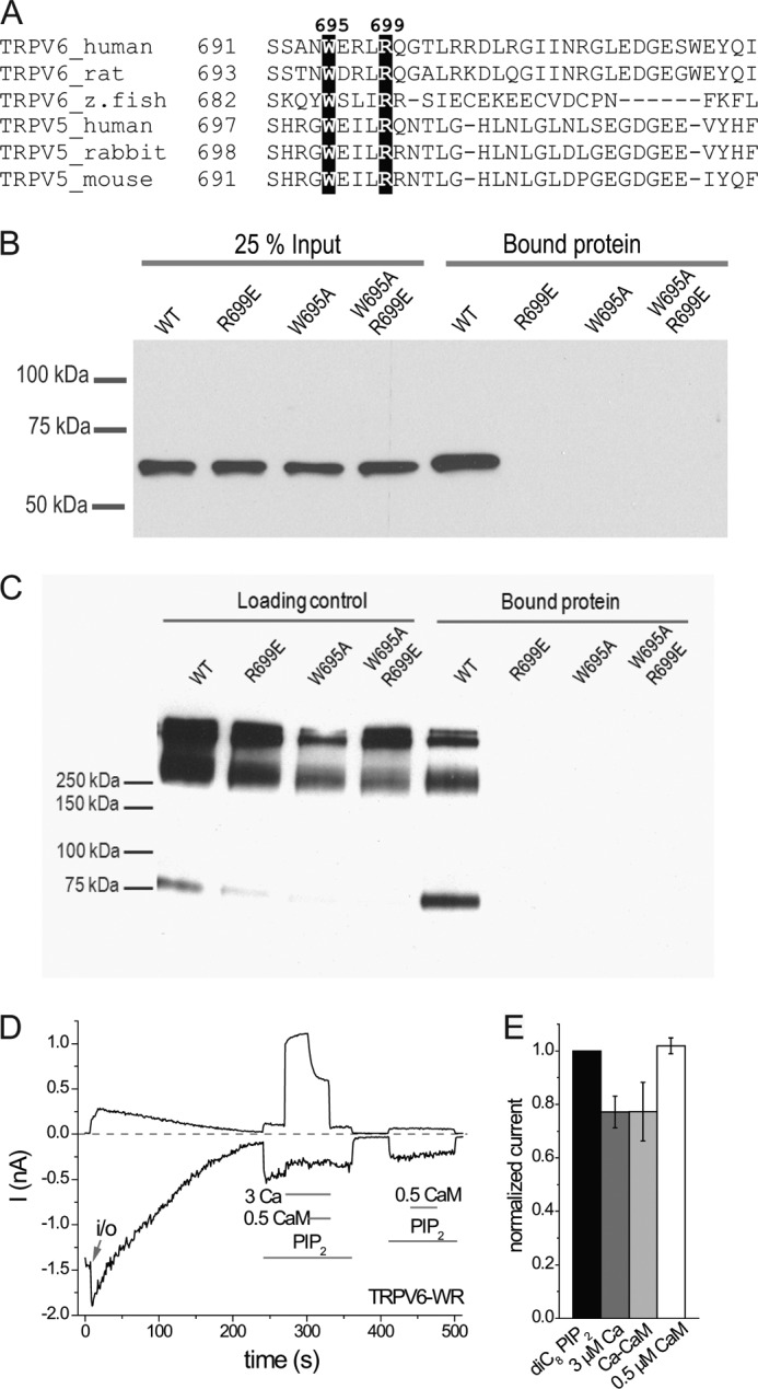
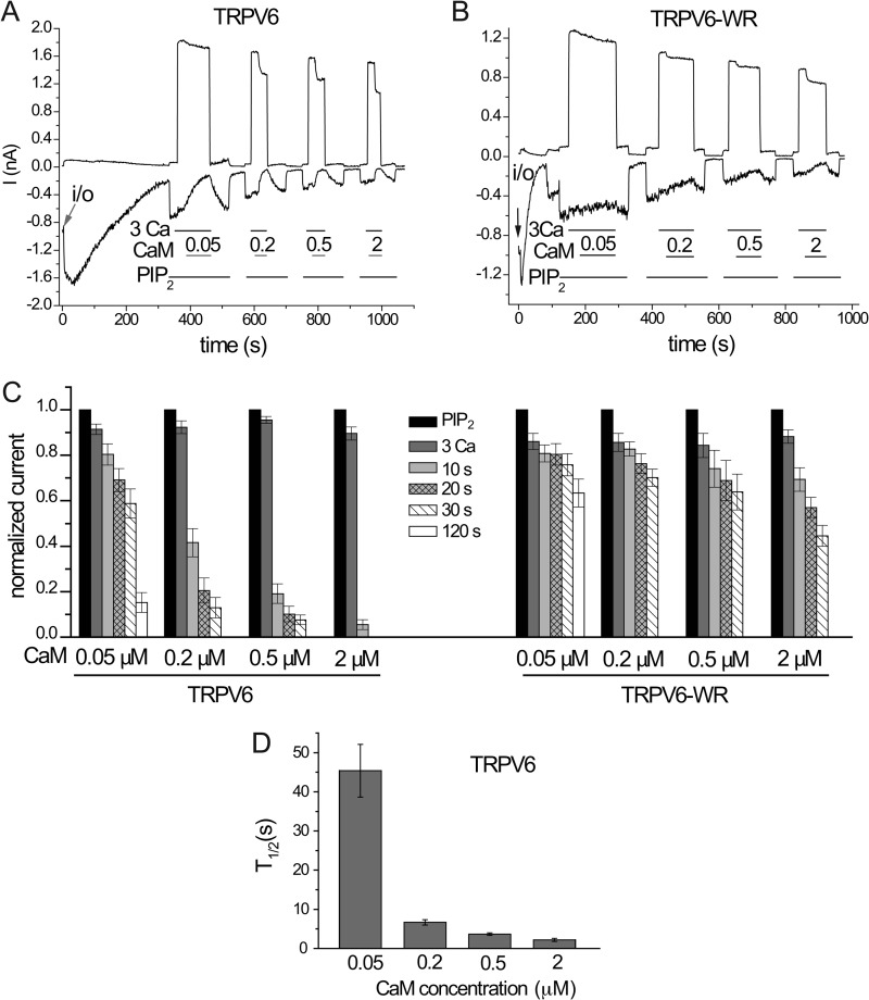
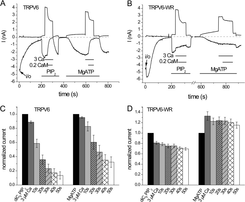
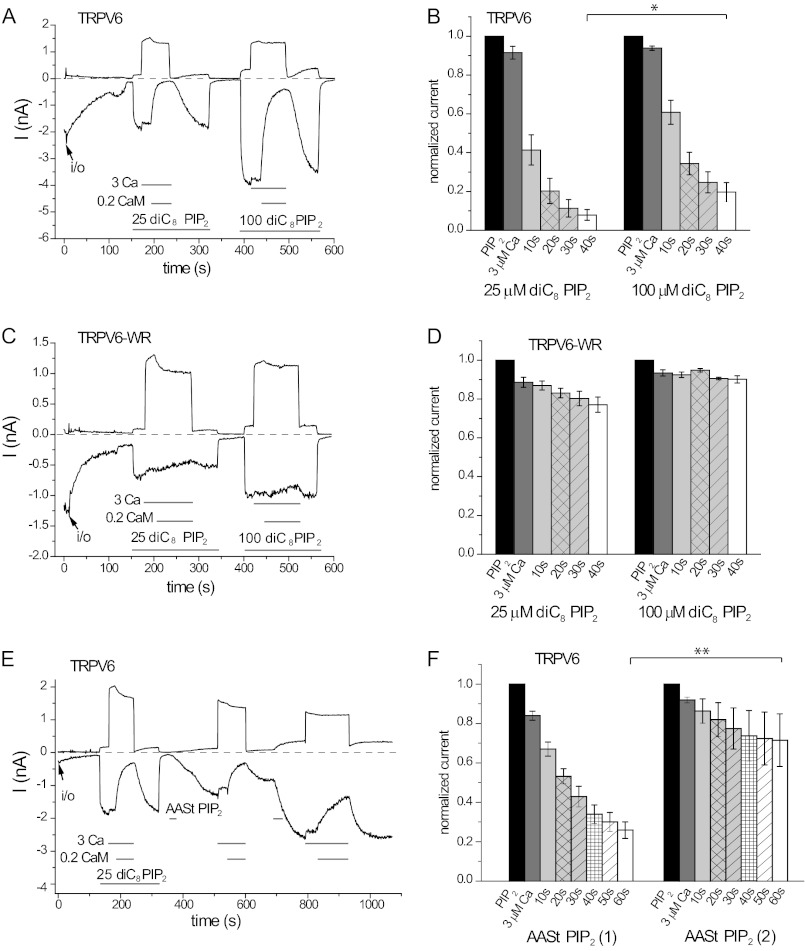
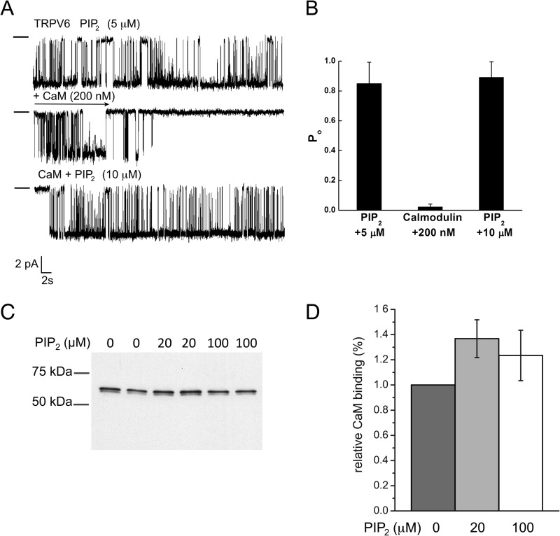
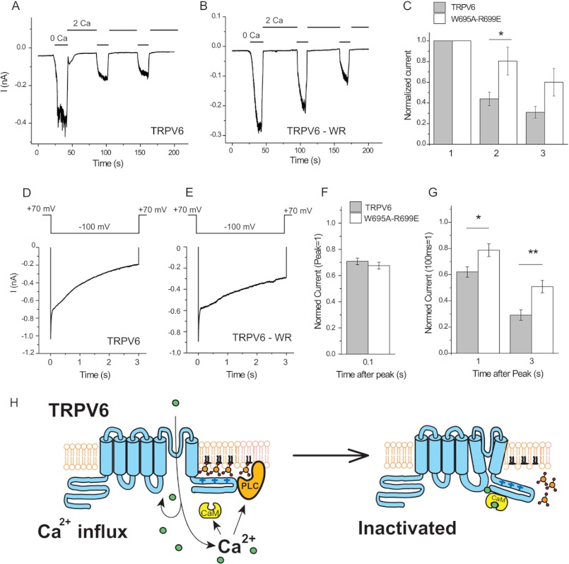
The epithelial Ca(2+) channel transient receptor potential vanilloid 6 (TRPV6) undergoes Ca(2+)-induced inactivation that protects the cell from toxic Ca(2+) overload and may also limit intestinal Ca(2+) transport. To dissect the roles of individual signaling pathways in this phenomenon, we studied the effects of Ca(2+), calmodulin (CaM), and phosphatidylinositol 4,5-bisphosphate (PI(4,5)P(2)) in excised inside-out patches. The activity of TRPV6 strictly depended on the presence of PI(4,5)P(2), and Ca(2+)-CaM inhibited the channel at physiologically relevant concentrations. Ca(2+) alone also inhibited TRPV6 at high concentrations (IC(50) = ∼20 μM). A double mutation in the distal C-terminal CaM-binding site of TRPV6 (W695A/R699E) essentially eliminated inhibition by CaM in excised patches. In whole cell patch clamp experiments, this mutation reduced but did not eliminate Ca(2+)-induced inactivation. Providing excess PI(4,5)P(2) reduced the inhibition by CaM in excised patches and in planar lipid bilayers, but PI(4,5)P(2) did not inhibit binding of CaM to the C terminus of the channel. Overall, our data show a complex interplay between CaM and PI(4,5)P(2) and show that Ca(2+), CaM, and the depletion of PI(4,5)P(2) all contribute to inactivation of TRPV6.


Figures

FIGURE 1.
FIGURE 1.
Ca2+-CaM inhibits TRPV6 activity in excised inside-out macropatches. Measurements on TRPV6-expressing and noninjected oocytes were performed as described under “Experimental Procedures.” Currents are shown at −103 mV (lower traces, Li+ current through TRPV6) and at +100 mV (upper traces, Cl current through the endogenous Ca2+-activated Cl channels). A, representative current traces in response to 3 μm Ca2+ in a noninjected oocyte. B, representative trace for a TRPV6-expressing oocyte, the applications of 25 μm diC8 PI(4,5)P2, 3 μm Ca2+, 0.5 μm CaM in the presence of 3 μm Ca2+, and 0.5 μm CaM alone are indicated by the horizontal lines. C, summary of the effects of Ca2+, Ca2+-CaM, and CaM in the absence of Ca2+ (n = 6). D, representative traces for the effect Ca2+-CaM in a patch where rundown of TRPV6 currents was very slow. E, summary of the time course of inhibition without application of exogenous PI(4,5)P2 (endo PIP2) and on currents stimulated by diC8 PI(4,5)P2 from five experiments.
FIGURE 2.
FIGURE 2.
Concentration dependence of the Ca2+ inhibition of TRPV6 activity in excised inside-out macropatches. A, representative trace for the application of 3, 30, and 100 μm free Ca2+ on wild-type TRPV6 activity induced by 25 μm diC8 PI(4,5)P2. This experiment was performed in symmetrical Cl-free conditions, as described under “Experimental Procedures”; traces at −100 and +100 mV are shown. Note the absence of the Cl current. B, summary of the data (n = 5–18). The IC50 for Ca2+ inhibition is 20.8 μm, and the Hill coefficient is 1.01.
FIGURE 3.
FIGURE 3.
CaM binds to TRPV6 via a distal C-terminal binding site. A, proposed CaM-binding sites in TRPV5 and TRPV6. The amino acid sequences correspond to the equivalent fragments in the human TRPV6. B, CaM-Sepharose pulldown assay using MBP fusion proteins of TRPV6 wild-type cytoplasmic C terminus (amino acids 579–725), truncated C terminus without the distal region (Δ694–725), and wild-type cytoplasmic N terminus (amino acids 1–326) in the presence of various Ca2+ concentrations or 2 mm EGTA. Bound proteins were detected by Western blot analysis using anti-MBP antibody. The images are representative of five or six experiments. C, CaM binding assays were performed using the crude membrane fractions from HEK293 cells expressing full-length wild-type Myc-hTRPV6 or mutant Myc-hTRPV6Δ694–725 in the presence of 20 μm Ca2+. Bound proteins eluted from the CaM beads were detected by Western blot analysis using an anti-Myc antibody. The images are representative of eight experiments.
FIGURE 4.
FIGURE 4.
Two highly conserved amino acid residues in the distal C terminus of TRPV6 are responsible for interacting with CaM. A, sequence alignment shows that residues Trp-695 and Arg-699 in hTRPV6 are fully conserved among all studied TRPV5 and TRPV6 species (16). B, CaM-Sepharose pulldown assays were performed in the presence of 100 μm Ca2+ on the isolated C terminus of wild-type and mutant TRPV6. The image is representative of five experiments. C, CaM-Sepharose pulldown assays were performed in the presence of 20 μm Ca2+ for full-length, wild-type, and three mutated TRPV6 proteins transiently expressed in HEK293 cells. Input proteins for loading control and bound proteins eluted from the CaM beads are detected by Western blot using an anti-Myc antibody. The images are representative of three experiments. D, representative trace for the effect of Ca2+, Ca2+-CaM, and CaM on double mutant W695A/R699E (TRPV6-WR) channel activity stimulated by 25 μm diC8 PI(4,5)P2 in excised inside-out macropatches. E, summary data for D normalized to the current evoked by diC8 PI(4,5)P2 (n = 5).
FIGURE 5.
FIGURE 5.
Concentration dependence of the effect of CaM on TRPV6 in excised inside-out macropatches. A and B, representative traces for the effects of different concentrations of CaM in the presence of 3 μm Ca2+ on wild-type TRPV6 (TRPV6) and double mutant W695A/R699E (TRPV6-WR) activated by 25 μm diC8 PI(4,5)P2. C, summary data, normalized to the current evoked by 25 μm diC8 PI(4,5)P2 (n = 5–8). D, T½ values for the different concentrations of CaM.
FIGURE 6.
FIGURE 6.
Ca2+-CaM inhibits TRPV6 activity stimulated by MgATP in excised inside-out patches. A and B, representative traces for the application of Ca2+ (3 μm) and Ca2+-CaM (3 μm Ca2+ and 0.2 μm CaM) on wild-type TRPV6 and the W695A/R699E double mutant TRPV6 (TRPV6-WR) activity, respectively. Channel activity was stimulated by 25 μm diC8 PI(4,5)P2 and 2 mm MgATP (2 mm NaATP and 2 mm Mg2+). C and D, summary of the data for six or seven experiments.
FIGURE 7.
FIGURE 7.
PI(4,5)P2 competes with Ca2+-CaM on TRPV6 activity in excised inside-out patches. A, representative trace for the effect of 3 μm Ca2+ and Ca2+-CaM (3 μm Ca2+ and 0.2 μm CaM) on wild-type TRPV6, in the presence of 25 and 100 μm diC8 PI(4,5)P2. B, summary data for A, normalized to the current evoked by diC8 PI(4,5)P2 with the corresponding concentration for the wild-type TRPV6 channel. The inhibition by CaM at 40 s is significantly less at 100 μm diC8 PI(4,5)P2 than at 25 μm (n = 9, p = 0.0104). C, representative experiment for the double mutant TRPV6 (TRPV6-WR). D, summary for the effect of CaM on the W695A/R699E mutant of TRPV6 (n = 4). E, representative trace for the effect of 3 μm Ca2+ and 0.2 μm Ca2+-CaM on wild-type TRPV6 activity, in the presence of natural AASt PI(4,5)P2 (10 μm). Because of the micelle form of AASt PI(4,5)P2 in the bath solution, the amount of AASt PI(4,5)P2 incorporated into the patch membrane was associated with the time of application. F, summary data for E, normalized to the current evoked by AASt PI(4,5)P2. The inhibition by CaM was significantly less at the second application of AASt PI(4,5)P2 than at the first one, when measured at 60 s (n = 6, p = 0.0056). Asterisks denote statistical significance.
FIGURE 8.
FIGURE 8.
PI(4,5)P2 competes with CaM in planar lipid bilayers, but not in biochemical binding experiments. A, planar lipid bilayer measurements were performed as described under “Experimental Procedures.” TRPV6 activity was stimulated by 5 μm diC8 PI(4,5)P2. Then 200 nm CaM was applied, and PI(4,5)P2 concentration was increased to 10 μm. Clamping potential was −100 mV. The closed state is indicated by a horizontal line on the left side of traces. B, summary of the bilayer experiments (n = 5 for CaM inhibition and n = 3 for PI(4,5)P2 reactivation). C, CaM-Sepharose pulldown assay was performed using MBP fusion proteins of TRPV6 wild-type C terminus in the absence and the presence of 20 or 100 μm diC8 PI(4,5)P2 as described under “Experimental Procedures.” Free Ca2+ concentration was 100 μm. Bound proteins were detected by SDS-PAGE and Western-blotting using anti-MBP antibodies. D, statistical summary based on five measurements.
FIGURE 9.
FIGURE 9.
Ca2+-induced inactivation is reduced but not eliminated in the W695A/R699E mutant. Whole cell patch clamp experiments in TRPV6-expressing HEK cells were performed as described under “Experimental Procedures.” A and B, representative measurements for wild-type and W695A/R699E (WR) TRPV6 channels at constant −60 mV holding potential. At the beginning of the measurements, the cells were kept in a nominally Ca2+-free solution containing 1 mm Mg2+. Monovalent currents were evoked by the application of 2 mm EGTA in bivalent free extracellular solution (0 Ca); then Ca2+-induced inactivation was induced by the application of a solution containing 2 mm Ca2+ and no Mg2+ (2 Ca). C, summary of current amplitudes in the first, second, and third applications of EGTA. The difference between wild-type and the mutant channel was statistically significant in the second (p = 0.024, n = 11), but not at the third pulse (p = 0.058). D and E, Ca2+-induced inactivation of Ca2+ currents for wild-type and mutant TRPV6 channels. Ca2+ currents were initiated by a 3-s voltage step from +70 to −100 mV in an extracellular solution containing 10 mm Ca2+. The traces shown are averages for six measurements for both groups. F, summary of inactivation kinetics at 100 ms; the data are normalized to the point 3 ms after the voltage step, to avoid capacitative artifacts. G, summary at 1 and 3 s after the −100 mV voltage step; the data are normalized to the current 100 ms after the voltage step, after the initial fast phase inactivation. The difference between wild-type and mutant channel was statistically significant both at 1 s (p = 0.025) and at 3 s (p = 0.0054). Asterisks denote statistical significance. H, model for Ca2+-induced inactivation of TRPV6.

Similar articles

Cited by

References

    1. Hoenderop J. G., Nilius B., Bindels R. J. (2005) Calcium absorption across epithelia. Physiol. Rev. 85, 373–422 - PubMed
    1. Weissgerber P., Kriebs U., Tsvilovskyy V., Olausson J., Kretz O., Stoerger C., Vennekens R., Wissenbach U., Middendorff R., Flockerzi V., Freichel M. (2011) Male fertility depends on Ca2+ absorption by TRPV6 in epididymal epithelia. Sci. Signal. 4, ra27. - PubMed
    1. Nilius B., Prenen J., Hoenderop J. G., Vennekens R., Hoefs S., Weidema A. F., Droogmans G., Bindels R. J. (2002) Fast and slow inactivation kinetics of the Ca2+ channels ECaC1 and ECaC2 (TRPV5 and TRPV6). Role of the intracellular loop located between transmembrane segments 2 and 3. J. Biol. Chem. 277, 30852–30858 - PubMed
    1. Thyagarajan B., Benn B. S., Christakos S., Rohacs T. (2009) Phospholipase C-mediated regulation of transient receptor potential vanilloid 6 channels. Implications in active intestinal Ca2+ transport. Mol. Pharmacol. 75, 608–616 - PMC - PubMed
    1. Suh B. C., Hille B. (2008) PIP2 is a necessary cofactor for ion channel function. How and why? Annu. Rev. Biophys. 37, 175–195 - PMC - PubMed

Publication types

MeSH terms