. 2012;7(12):e52166.
doi: 10.1371/journal.pone.0052166. Epub 2012 Dec 31.

Transcriptional program of ciliated epithelial cells reveals new cilium and centrosome components and links to human disease

Affiliations

Transcriptional program of ciliated epithelial cells reveals new cilium and centrosome components and links to human disease

Ramona A Hoh et al. PLoS One. 2012.

Abstract

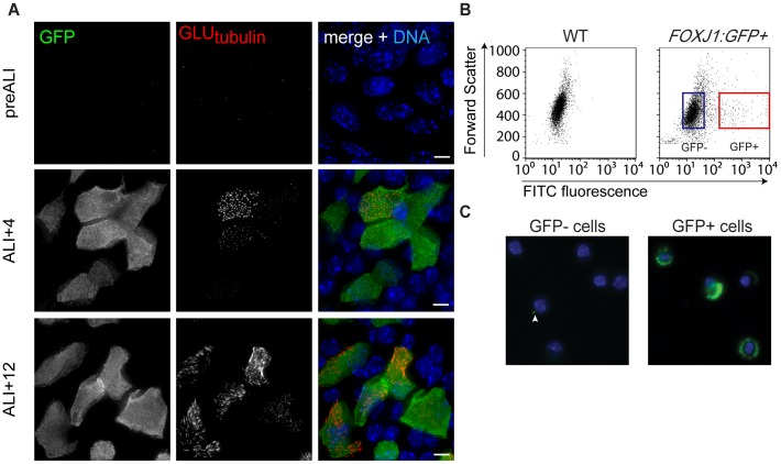
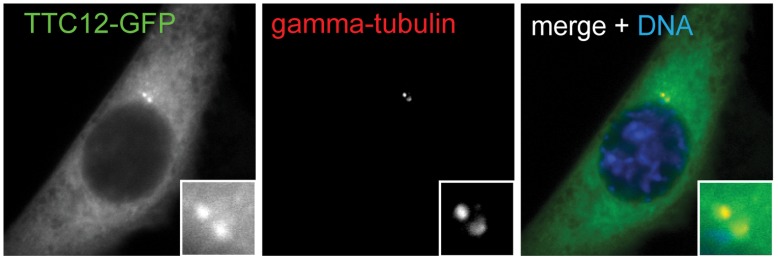
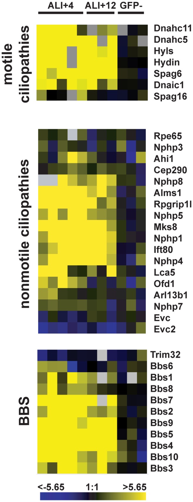
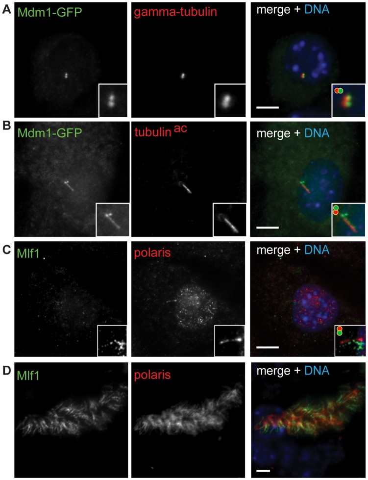
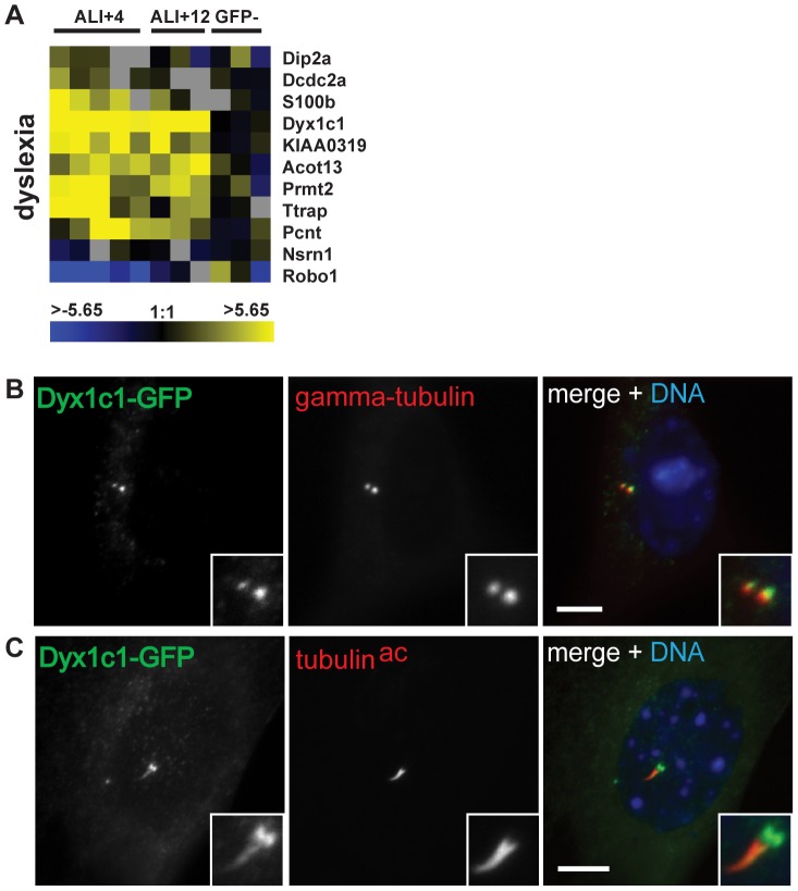
Defects in the centrosome and cilium are associated with a set of human diseases having diverse phenotypes. To further characterize the components that define the function of these organelles we determined the transcriptional profile of multiciliated tracheal epithelial cells. Cultures of mouse tracheal epithelial cells undergoing differentiation in vitro were derived from mice expressing GFP from the ciliated-cell specific FOXJ1 promoter (FOXJ1:GFP). The transcriptional profile of ciliating GFP+ cells from these cultures was defined at an early and a late time point during differentiation and was refined by subtraction of the profile of the non-ciliated GFP- cells. We identified 649 genes upregulated early, when most cells were forming basal bodies, and 73 genes genes upregulated late, when most cells were fully ciliated. Most, but not all, of known centrosome proteins are transcriptionally upregulated early, particularly Plk4, a master regulator of centriole formation. We found that three genes associated with human disease states, Mdm1, Mlf1, and Dyx1c1, are upregulated during ciliogenesis and localize to centrioles and cilia. This transcriptome for mammalian multiciliated epithelial cells identifies new candidate centrosome and cilia proteins, highlights similarities between components of motile and primary cilia, and identifies new links between cilia proteins and human disease.


Conflict of interest statement

Competing Interests: The authors have declared that no competing interests exist.

Figures

Figure 1
Figure 1. Microarray analysis of ciliating mouse tracheal epithelial cells. (
A) Immunofluorescence staining showing representative images of GFP expression and ciliogenesis in FOXJ1:GFP MTEC cultures before establishment of the air-liquid interface (pre-ALI), at 4 days after ALI (ALI+4) and at 12 days after ALI (ALI+12). MTEC cultures were stained with antibodies against GFP, against glutamylated tubulin to mark cilia and basal bodies, and with DAPI to mark nuclei. GFP is expressed from the FOXJ1 promoter early during ciliogenesis, but is not expressed pre-ALI. At ALI+4, most FOXJ1:GFP+ cells are undergoing centriole formation but have not formed cilia. At ALI+12, most FOXJ1:GFP+ cells are forming cilia or have completed ciliogenesis. Scale bars, 5 µm. (B) FACS analysis of cells dissociated from a trachea from a wild type mouse (left panel) and a trachea from a FOXJ1:GFP-expressing mouse (right panel). The red and blue rectangles are representative gates used to sort GFP+ and GFP- populations, respectively. (C) Sorted cell populations were stained with DAPI (blue) and combined acetylated alpha-tubulin and gamma-tubulin antibodies (green) to detect cilia and basal bodies. After sorting, 90–95% of cells in the GFP+ population at ALI+12 had observable cilia and or amplified basal bodies, whereas <1% of cells in the GFP- population stained positive for these markers, although some had a single primary cilium (arrowhead).
Figure 2
Figure 2. Subtractive analysis of MTEC microarray data.
(A) Experimental flowchart detailing microarray analysis. (B) The number of differentially expressed genes in ciliated MTECs at adj. p-values <0.05, <0.01 and <0.001 before subtraction (non-subtracted) and after subtraction. The non-subtracted gene list is the transcriptional profile of GFP+ cells compared to a universal mouse reference RNA. The subtracted gene list was derived analytically by comparing the transcriptional profile of GFP+ cells to that of GFP- cells from MTEC cultures. The number of genes that show 2-fold or greater differential expression and have adjusted p-values of 0.05, 0.01 and 0.001 are shown before and after subtractive analysis. After subtraction, 649 genes were significantly upregulated at ALI+4 with respect to GFP- cells, and 73 genes at ALI+12. 143 genes were downregulated at ALI+4 and none at ALI+12 (adj. p<0.05). (C) The overlap of significantly upregulated and downregulated genes (adj. p<0.05) from ALI+4, ALI+12 and GFP- pools decreases after subtraction. The GFP- group in both panels represents genes that are differentially expressed in non-ciliated cells relative to the universal reference RNA. M = log2(fold change).
Figure 3
Figure 3. Functional mapping of candidate centrosome/cilia proteins and identification of novel centrosome components.
Candidate genes were organized by known physical and genetic interactions mined from Mitocheck, the Human Protein Reference Database and BioGrid. Clusters containing genes associated with cell cycle control (A), hedgehog signaling (B) and the PCM1 complex (C) are shown.
Figure 4
Figure 4. Genes encoding centrosome proteins are differentially regulated during basal body formation.
(A) Upregulation of regulatory and structural proteins required for centriole duplication in cycling cells and (B) heat map of putative and known centrosomal components. Genes are shown in rows and replicate arrays are shown in columns. Three biological replicates were performed for ALI+4, ALI+12 and GFP-. In addition, two technical replicates were performed for two of the ALI+4 biological replicates (columns 1,2 and 4,5) for a total of five columns. Data were zero-transformed against non-ciliated (GFP-) cells. The scale indicates the fold change in expression (log2). Grey boxes represent gene spots that failed to pass quality control filters for the indicated array.
Figure 5
Figure 5. Identification of a novel centrosome component.
NIH/3T3 cells were transfected with a plasmid encoding TTC12-GFP. TTC12-GFP localizes to the centrosome as shown by overlap with gamma-tubulin staining. Green, TTC12-GFP. Red, gamma-tubulin.
Figure 6
Figure 6. Differential regulation of disease genes associated with motile and nonmotile ciliopathies.
Heat maps showing the expression of genes associated with motile (top) and nonmotile (middle) ciliopathies, as well as genes linked to Bardet-Biedl syndrome (bottom), a model ciliopathy.
Figure 7
Figure 7. Identification of new links between centrosomes, cilia and human disease.
(A, B) NIH/3T3 cells were transfected with a plasmid encoding MDM1-GFP. MDM1-GFP localizes to centrosomes (A) and the primary cilium (B). NIH/3T3 cells and MTECs were stained for MLF1 with an anti-MLF1 antibody. MLF1 localizes to the primary cilium in NIH/3T3 cells (C) and motile cilia in MTECs (D). Insets show higher magnification of merged images, slightly offset as indicated by circles in upper left. Scale bars, 5 µm. Green, GFP (A, B); MLF1 (C, D). Red, gamma-tubulin (A); acetylated tubulin (B); polaris (C, D).
Figure 8
Figure 8. A dyslexia candidate disease gene is transcriptionally upregulated and localizes to the centrosome.
(A) Heat maps depicting the expression of dyslexia candidate genes in ciliated MTECs. Dyx1c1 is among the most highly upregulated genes, and Robo1 is strongly downregulated. (B-D) A DYX1C1-GFP fusion protein was expressed in NIH/3T3 cells. DYX1C1-GFP localizes to the centrosome (B) and in some cells the primary cilium (C). Insets show positively staining regions at higher magnification. Scale bars, 5 um. Green, GFP (B, C). Red, gamma-tubulin (B); acetylated tubulin (C).

References

    1. Vincensini L, Blisnick T, Bastin P (2011) 1001 model organisms to study cilia and flagella. Biol Cell 103: 109–130. - PubMed
    1. Berbari NF, O’Connor AK, Haycraft CJ, Yoder BK (2009) The primary cilium as a complex signaling center. Curr Biol 19: R526–535. - PMC - PubMed
    1. Goetz SC, Anderson KV (2010) The primary cilium: a signalling centre during vertebrate development. Nat Rev Genet 11: 331–344. - PMC - PubMed
    1. Fisch C, Dupuis-Williams P (2011) Ultrastructure of cilia and flagella - back to the future! Biol Cell. 103: 249–270. - PubMed
    1. Christensen ST, Guerra CF, Awan A, Wheatley DN, Satir P (2003) Insulin receptor-like proteins in Tetrahymena thermophila ciliary membranes. Curr Biol 13: R50–52. - PubMed

Publication types

MeSH terms