. 2012;7(12):e52278.
doi: 10.1371/journal.pone.0052278. Epub 2012 Dec 27.

Horizontal gene transfer contributed to the evolution of extracellular surface structures: the freshwater polyp Hydra is covered by a complex fibrous cuticle containing glycosaminoglycans and proteins of the PPOD and SWT (sweet tooth) families

Affiliations

Horizontal gene transfer contributed to the evolution of extracellular surface structures: the freshwater polyp Hydra is covered by a complex fibrous cuticle containing glycosaminoglycans and proteins of the PPOD and SWT (sweet tooth) families

Angelika Böttger et al. PLoS One. 2012.

Abstract

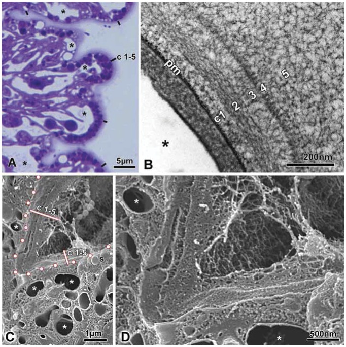
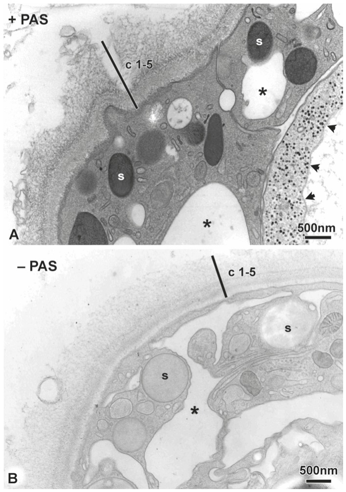
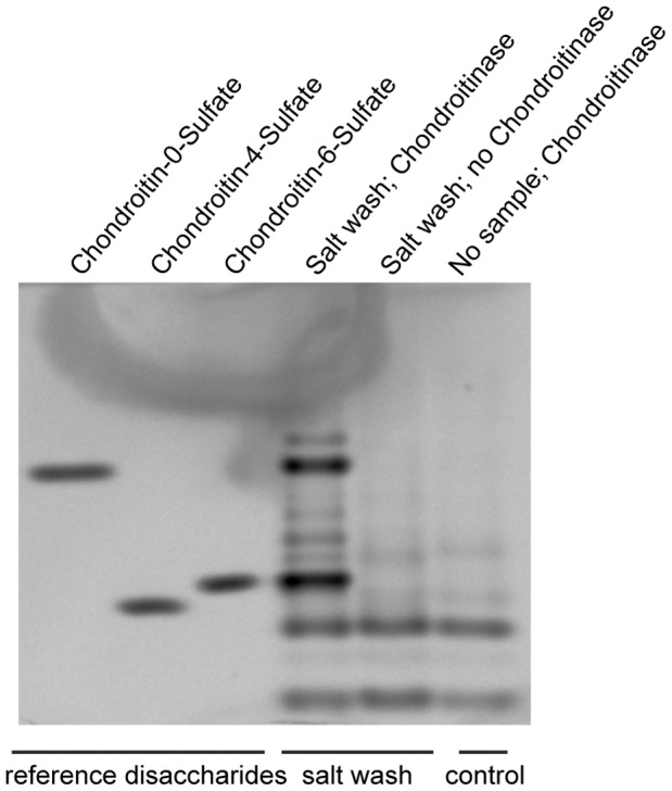
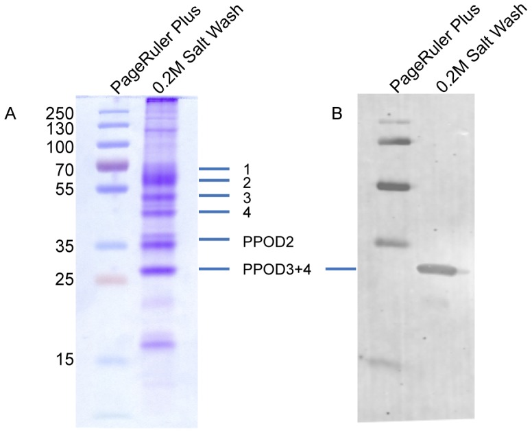
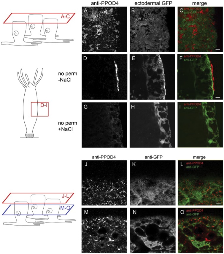
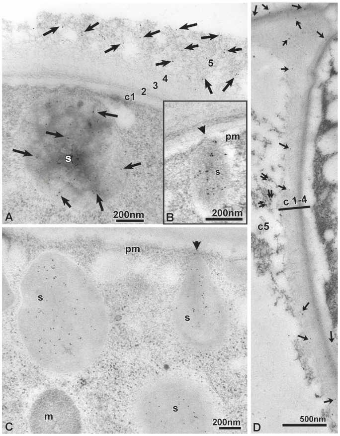
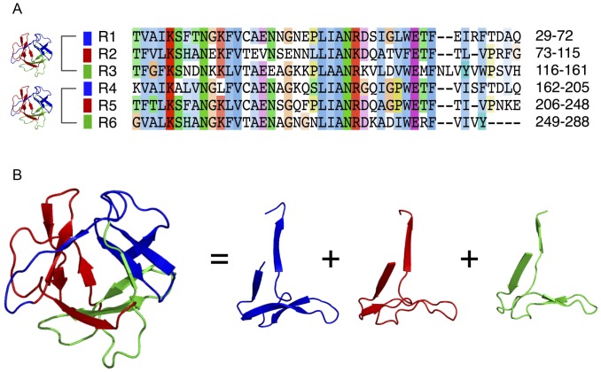
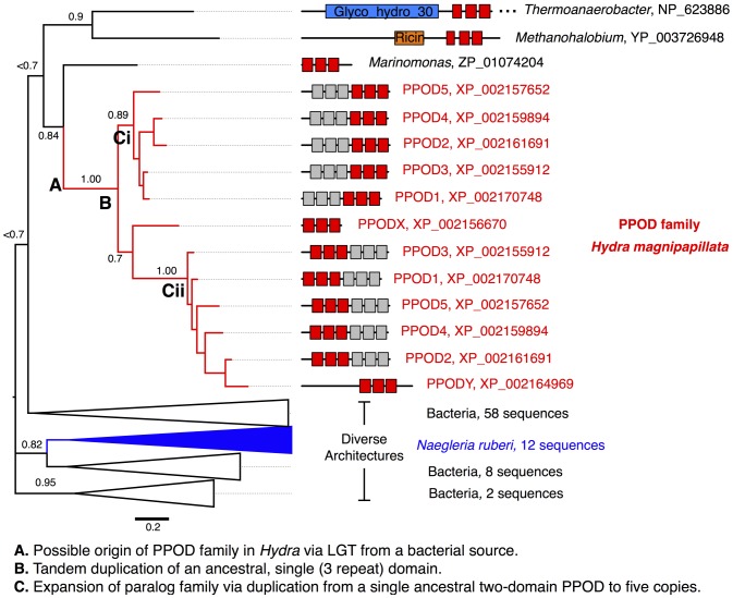
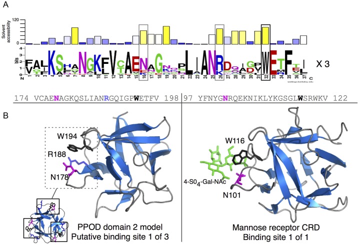
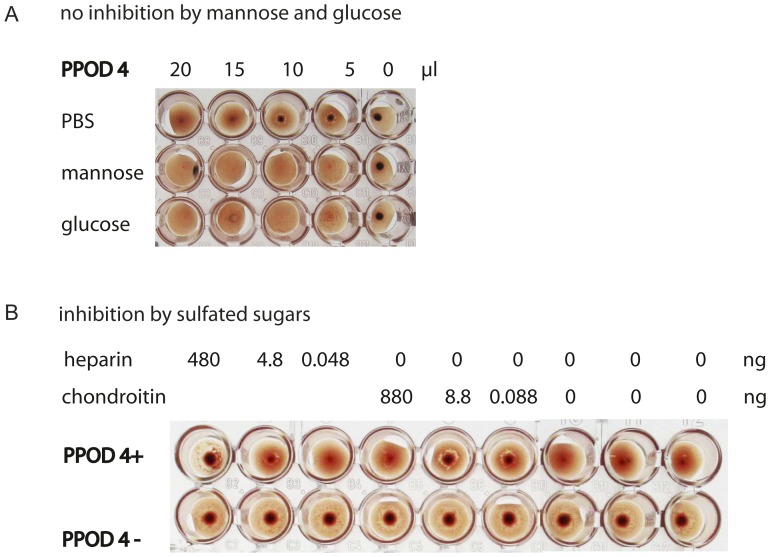
The single-cell layered ectoderm of the fresh water polyp Hydra fulfills the function of an epidermis by protecting the animals from the surrounding medium. Its outer surface is covered by a fibrous structure termed the cuticle layer, with similarity to the extracellular surface coats of mammalian epithelia. In this paper we have identified molecular components of the cuticle. We show that its outermost layer contains glycoproteins and glycosaminoglycans and we have identified chondroitin and chondroitin-6-sulfate chains. In a search for proteins that could be involved in organising this structure we found PPOD proteins and several members of a protein family containing only SWT (sweet tooth) domains. Structural analyses indicate that PPODs consist of two tandem β-trefoil domains with similarity to carbohydrate-binding sites found in lectins. Experimental evidence confirmed that PPODs can bind sulfated glycans and are secreted into the cuticle layer from granules localized under the apical surface of the ectodermal epithelial cells. PPODs are taxon-specific proteins which appear to have entered the Hydra genome by horizontal gene transfer from bacteria. Their acquisition at the time Hydra evolved from a marine ancestor may have been critical for the transition to the freshwater environment.


Conflict of interest statement

Competing Interests: The authors have declared that no competing interests exist.

Figures

Figure 1
Figure 1. Light and electron microsopic images of cryo-fixed Hydra cuticle.
A: Overview of 0.5 µm semi-thin Epon section stained with the basic dye toluidine blue. The cuticle (c1–5) covering the whole H. vulgaris polyp is clearly visible. Asterisks mark the pleiomorphic intra−/intercellular vacuolar system. Scale bar: 5 µm. B: Transmission EM reveals the fibrous appearance of layer c2 and the periodically arranged rod-like substructures forming layer c4. Plasma membrane marked by (pm), asterisk marks a vacuole. Note the strong contrast of all 5 cuticle layers and the plasma membrane obtained with the PAS-reagent (see also Fig. 2). Scale bar: 200 nm. C: Scanning EM of freeze-fractured H. magnipapillata ectodermal epithelium reveals the overlying cuticle (c1–5) as a bulky structure (note: individual cuticle layers cannot be distinguished in the SEM-samples). Dots mark the apical plasma membrane of the epithelium, asteriks mark the vacuolar system, secretory granules are indicated by “s”. Scale bar: 1 µm. D: Magnified view of Fig. 1C. Scale bar: 500 nm.
Figure 2
Figure 2. PAS cytochemistry of Hydra cuticle.
Periodic acid-thiocarbohydrazide-silver proteinate staining was performed on Epon sections from H. vulgaris. A: Cuticle layers c1–5 react positively as do apical secretory granules (s) and glycogen particles (arrow-heads) in a neighbouring nematocyst; the asterisk marks a vacuole. Scale bar: 500 nm. B: Negative control for the PAS-reaction (omission of periodic acid oxidation); faint unspecific staining results from binding of thiocarbohydrazide to the osmium tetroxide used for freeze-substitution. Scale bar: 500 nm.
Figure 3
Figure 3. Chondroitinsulfates in hypertonic salt wash.
The salt wash was digested with chondroitinase and the resulting disaccharides labeled with AMAC and separated by PAGEFS. The left three lanes are standard disaccharides.
Figure 4
Figure 4. Soluble protein content of hypertonic salt wash.
A: SDS-PAGE gel of 0.2 M NaCl wash stained with coomassie. Major bands were excised and identified by mass spectrometry. Positions of the PPOD 2, 3 and 4 bands are indicated. Bands 1–4 represent members of SWT protein family. See text for details. B: Immunoblot for this gel stained with anti-PPOD4 antibody.
Figure 5
Figure 5. Immunofluorescence staining of Hydra polyps with anti-PPOD4 antibody.
Polyps were fixed with PFA (A-I) or Lawdowsky’s fixative (J–O), stained with anti-PPOD4 or anti-GFP antibody and examined in the confocal microscope. Schematic diagrams indicate the positions of the optical sections shown. Polyps in A-I were not permeabilized and show staining of the extracellular surface. Polyps in J-O were permeabilized to permit staining of intracellular vesicles. See text for details.
Figure 6
Figure 6. Immuno-EM localisation of PPOD.
A: Immunogold labeling of PPOD (visualised by anti-chicken 10 nm colloidal gold) in the cuticle layer c5 and in subapical secretory granules (s) of ectodermal epithelial cells. Freeze-substitution with pure acetone followed by LR-white embedding. Scale bar: 200 nm. B: PPOD-positive secretory granule (s) in contact (arrow-head) with the plasma membrane (pm). Scale bar: 200 nm. C: Subapical secretory granules (s) are all PPOD-positive. A mitochondrium is lettered with (m), the plasma membrane with (pm). Scale bar: 200 nm. D: Scarce PPOD-immunogold-labelling can also be seen throughout cuticle layers c2–4 (marked by arrows), in addition to PPOD-labelling of layer c5 (double arrows). Scale bar: 500 nm.
Figure 7
Figure 7. Internal repeats and three-dimensional structure of Hydra PPOD.
A) Alignment of six internal repeats detected within the PPOD4 sequence using the RADAR algorithm. B) Structural model of a single β-trefoil domain in PPOD4 as inferred by Phyre. Three internal sequence repeats (coloured ribbon models) correspond to three repeated supersecondary structures that form a single β-trefoil fold.
Figure 8
Figure 8. Evolutionary analysis of Hydra PPOD reveals a link to bacterial carbohydrate-binding proteins.
A bayesian inference phylogenetic tree was generated for the Hydra PPOD family and the top-scoring related sequences retrieved by BLAST. The Hydra PPOD family forms a single clade restricted to Hydra and has expanded by tandem duplication of a single β-trefoil domain, followed by whole-gene duplications. The closest ancestral sequences are of bacterial origin. The most related sequences include additional domains such as glycosyl hydrolase domains and sugar-binding domains (e.g., ricin). Unlike most of the detected bacterial homologs, the closest outgroup of the Hydra PPOD clade (a sequence from Marinomonas) has a single β-trefoil domain, supporting its close relationship to the inferred ancstral PPOD domain. Posterior probabilities are indicated above the major clades. See Fig. S4 for an expanded version of this tree.
Figure 9
Figure 9. Structural modeling suggests a carbohydrate-binding function for PPOD.
A) A sequence logo was generated by combining all repeats from the PPOD family into a single alignment. Above the sequence logo, the solvent-accessibility has been plotted as calculated from the binding-site associated repeat shown in (B). B) Left - a structural model of PPOD4 domain 2 with putative sugar-binding Trp residues highlighted in red. Right - Structure of the CRD from the mannose receptor bound 4-SO4-Gal-NAc (PDB ID 1FWV). The matching N and W residues are indicated in the alignment above, as well as an additional conserved residue (R188) that is also located close to the putative binding site.
Figure 10
Figure 10. PPOD agglutinates erythrocytes.
Haemagglutination assays with rabbit erythrocytes. A: Addition of increasing amounts PPOD4 protein agglutinates erythrocytes and prevents their sedimentation (dark dot at 0 µl PPOD4 indicates sedimentation of erythrocytes, which are not agglutinated). Addition of mannose or glucose does not prevent PPOD4 induced agglutination. B: Erythrocytes sediment in the absence of PPOD4 protein (lower row). Addition of PPOD4 prevents this due to agglutination. Addition of heparin or chondroitin (GalNAc-4-SO4) to PPOD prevents PPOD4 induced agglutination in a concentration dependent manner.

References

    1. Sarras MP, Deutzmann R (2001) Hydra and Niccolo Paganini (1782–1840)–two peas in a pod? The molecular basis of extracellular matrix structure in the invertebrate, Hydra . Bioessays 23: 716–724. - PubMed
    1. Lentz TL (1966) The Cell Biology of Hydra. Amsterdam: North-Holland publishing company. 199 p.
    1. Holtmann M, Thurm U (2001) Variations of concentric hair cells in a Cnidarian sensory epithelium (Coryne tubulosa). J Comp Neurol 432: 550–563. - PubMed
    1. Thomas MB, Edwards NC, Higgins RP (1995) Cryptohydra thieli n.gen., sp.: a meiofaunal marine hydroid (Hydroida, Athecata, Capitata). Invertebrate Biology 114: 107–118.
    1. Kuznetsov SG, Anton-Erxleben F, Bosch TC (2002) Epithelial interactions in Hydra: apoptosis in interspecies grafts is induced by detachment from the extracellular matrix. J Exp Biol 205: 3809–3817. - PubMed

Publication types

MeSH terms

Substances