. 2012;7(12):e52332.
doi: 10.1371/journal.pone.0052332. Epub 2012 Dec 31.

IL-13 promotes collagen accumulation in Crohn's disease fibrosis by down-regulation of fibroblast MMP synthesis: a role for innate lymphoid cells?

Affiliations

IL-13 promotes collagen accumulation in Crohn's disease fibrosis by down-regulation of fibroblast MMP synthesis: a role for innate lymphoid cells?

Jennifer R Bailey et al. PLoS One. 2012.

Abstract

Background: Fibrosis is a serious consequence of Crohn's disease (CD), often necessitating surgical resection. We examined the hypothesis that IL-13 may promote collagen accumulation within the CD muscle microenvironment.

Methods: Factors potentially modulating collagen deposition were examined in intestinal tissue samples from fibrotic (f) CD and compared with cancer control (C), ulcerative colitis (UC) and uninvolved (u) CD. Mechanisms attributable to IL-13 were analysed using cell lines derived from uninvolved muscle tissue and tissue explants.

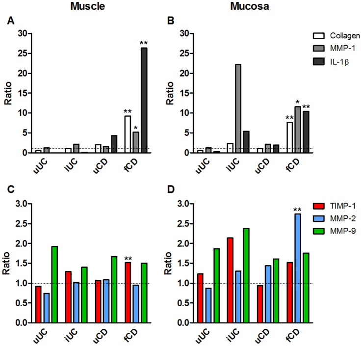
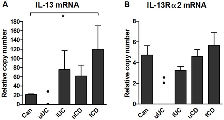
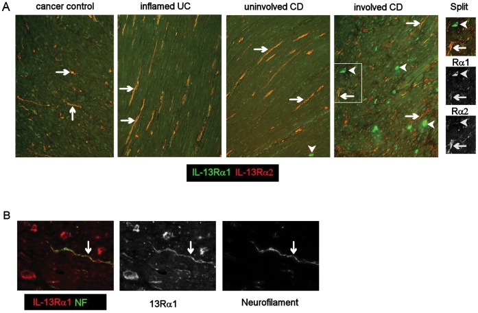
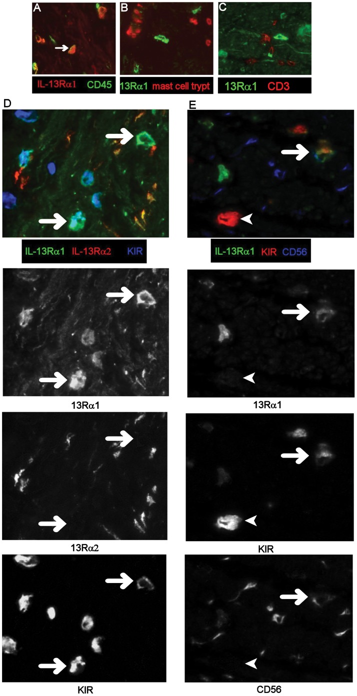
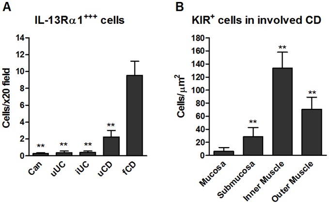
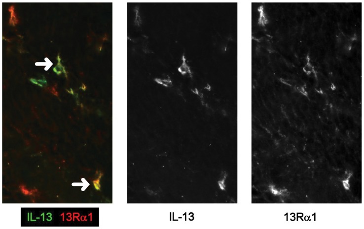
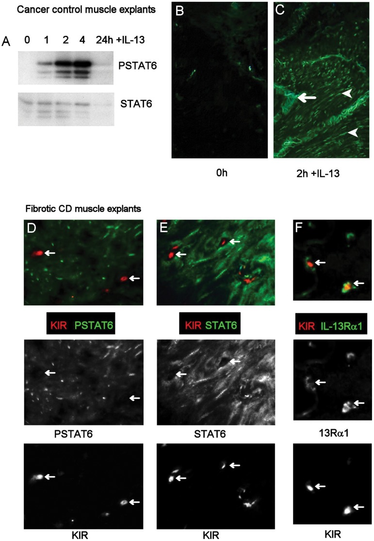
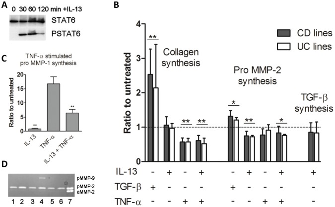
Results: In fCD muscle extracts, collagen synthesis was significantly increased compared to other groups, but MMP-2 was not co-ordinately increased. IL-13 transcripts were highest in fCD muscle compared to muscle from other groups. IL-13 receptor (R) α1 was expressed by intestinal muscle smooth muscle, nerve and KIR(+) cells. Fibroblasts from intestinal muscle expressed Rα1, phosphorylated STAT6 in response to IL-13, and subsequently down-regulated MMP-2 and TNF-α-induced MMP-1 and MMP-9 synthesis. Cells with the phenotype KIR(+)CD45(+)CD56(+/-)CD3(-) were significantly increased in fCD muscle compared to all other groups, expressed Rα1 and membrane IL-13, and transcribed high levels of IL-13. In explanted CD muscle, these cells did not phosphorylate STAT6 in response to exogenous IL-13.

Conclusions: The data indicate that in fibrotic intestinal muscle of Crohn's patients, the IL-13 pathway is stimulated, involving a novel population of infiltrating IL-13Rα1(+), KIR(+) innate lymphoid cells, producing IL-13 which inhibits fibroblast MMP synthesis. Consequently, matrix degradation is down-regulated and this leads to excessive collagen deposition.


Conflict of interest statement

Competing Interests: The authors have declared that no competing interests exist.

Figures

Figure 1
Figure 1. Increased collagen synthesis in muscle and submucosa in fibrotic CD tissue.
Full thickness sections of formalin fixed tissue stained with Haematoxylin van Geisens. Collagen is stained bright pink and nuclei are grey. (A) Cancer control (colon), (B) Inflamed UC (C) Uninvolved Crohn’s disease (terminal ileum) and (D) Crohn’s disease with stricture (terminal ileum) (stitched image). Arrows show dense collagen deposits in outer muscle layers. Dotted line defines the outer muscle/sub-mucosa (sm) border. Images captured with ×2.5 objective. Bars represent 1 mm.
Figure 2
Figure 2. Fibrogenesis is increased in CD muscle and mucosa tissues, without concomitant MMP-2 synthesis in muscle.
Tissue fragments were extracted and analysed by ELISA (collagen CICP synthesis, IL-1β, MMP-1, TIMP-1, TIMP-2) or zymography (MMP-2, MMP-9). Data are expressed as the ratio of the group mean for uUC, iUC, uCD or fCD to the mean for cancer control tissue for each parameter. Data are derived from 14–15 cancer controls, 3 uUC, 8 iUC, 8–12 uCD and 14–21 fCD patients. See Table S4 for actual means and SEM for each parameter. *p<0.05, **p<0.01 to cancer controls.
Figure 3
Figure 3. IL-13 transcription, but not IL-13Rα2 transcription, is increased fibrotic muscle.
(A) IL-13 mRNA and (B) IL-13 Rα2 mRNA in muscle tissue lysates. RNA was extracted from tissue fragments and processed for qPCR. Results were normalized to four stably expressed housekeeper genes. Data are derived from 14–15 cancer controls, 2 uUC, 8 iUC, 8–12 uCD and 14–21 fCD patients. Significant differences from fibrotic CD, *p<0.05.
Figure 4
Figure 4. IL-13 receptors are expressed on smooth muscle cells and neurofilaments in CD strictures.
Frozen sections were processed for double immunoflorescence. (Panel A) double labeling for IL-13 Rα1 and Rα2 in intestinal muscle tissue, arrows show double-stained cells, arrowheads show cells expressing Rα1 only at a very high level. Boxed area is shown as the colour image and its monochrome split. (B) Co-staining of IL-13Rα1 and neurofilament (NF) (green) and the monochrome split images. A,B, ×20 images.
Figure 5
Figure 5. Mononuclear cells expressing very high levels of IL-13Rα1 co-express CD45, KIR and CD56.
Images of frozen, immunostained tissue were captured using a 12 bit monochrome camera and images from the different colour channels combined to form two or three colour images. Multicoloured composites D and E, have been split into their original monochrome images to aid interpretation. Arrows show Rα1 co-localising with other markers. 13Rα1 co-localises with CD45 (A) but not mast cell tryptase (B) or CD3 (C). Panel D, 13Rα1 (green), 13Rα2 (red) and KIR (blue), plus split images for 13Rα1, 13Rα2 and KIR, note that 13Rα2 does not colocalise with 13Rα1+KIR+ cells (arrows). Panel E, 13Rα1 (green), KIR (red) and CD56 (blue). Split image shows a cell expressing all three markers (arrow), note there is also a strongly KIR-expressing cell (bottom left of image) which does not express 13Rα1 or CD56 A–C, ×40 images, D and E, ×64 images. Representative images from 8 patient samples.
Figure 6
Figure 6. IL-13Rα1 and KIR expressing-cells are increased in fibrotic CD, particularly in the muscle, by image analysis of immunostained frozen tissue sections.
(A) Total number of mononuclear cells/field expressing very high levels of Rα1 and no co-expression of Rα2 in muscle tissue (**p<0.01 to fCD), n = 8 for all groups except n = 3 for uUC. (B) Distribution of cells expressing very high levels of KIR in involved CD tissue. Data are the mean+SD of from 3 patients (20 images/patient), (*p<0.01 all comparisons).
Figure 7
Figure 7. IL-13 is co-localises with IL-13Rα1 in fCD muscle.
Double labeling, in frozen fibrotic CD tissue sections, for IL-13 (green) and IL-13Rα1 (red) and split monochrome images (×20).
Figure 8
Figure 8. IL-13 activates smooth muscle cells, but not KIR+ cells, in tissue explants.
Tissue explants were cultured with or without IL-13 for 1–24 h. Explants were then snap frozen and processed for Western blotting or immunoflorescence. (A–C) IL-13 signaling in explanted intestinal muscle tissue. (A) Time course for activation of STAT6 by Western blotting or (BC) by immunofluorescence in cancer control tissue. Arrowheads indicate positive nuclei, arrow indicates autoflorescence. (D–F) Double labeling for KIR and PSTAT6 (Panel D), STAT6 (Panel E), or Rα1 (Panel F) in fibrotic CD explanted tissue after 2 h incubation with IL-13. Arrows show KIR+ cells which colocalise with IL-13Rα1 but not with PSTAT6 or STAT6. A and C, ×64, B, ×20.
Figure 9
Figure 9. TGF-β drives co-ordinate collagen synthesis and pro-MMP-2 synthesis in intestinal muscle fibroblasts, whereas IL-13 has no effect on collagen synthesis and suppresses proMMP-2, in lines derived from uninvolved CD or UC tissue.
(A) Western blots showing phosphorylation of STAT6 by primary fibroblasts in response to exogenous IL-13. Representative blot from 6 experiments. (B) Effect of IL-13, TNF-α and/or TGF-β on collagen, MMP-2 or TGF-β synthesis in primary fibroblasts, Data represent the mean+SEM of the ratio of treated to untreated cultures from a minimum of 8 separate experiments. *p<0.05, **p<0.01 compared to untreated cultures. (C) Effect of IL-13 and/or TNF-α on pro-MMP-1 synthesis. Data represent the mean+SEM of the ratio of treated to untreated cultures from 8 separate experiments. **p<0.01 IL-13+TNF-α compared to TNF-α- or IL-13-stimulated cultures. (D) Zymogram showing the effect of IL-13, TNF-α and/or TGF-β on MMP-2 and MMP-9 synthesis (pMMP = pro MMP, aMMP = active MMP) (lane 1, untreated; lane 2, TGF-β; lane 3, IL-13; lane 4, TNF-α lane 5, TNF-α+IL-13; lane 6, TGF-β+IL-13; lane 7, MMP-2 standard).

References

    1. Burke JP, Mulsow JJ, O’Keane C, Docherty NG, Watson RW, et al. (2007) Fibrogenesis in Crohn’s disease. Am J Gastroenterol 102: 439–448. - PubMed
    1. Mourelle M, Salas A, Guarner F, Crespo E, Garcia-Lafuente A, et al. (1998) Stimulation of transforming growth factor beta1 by enteric bacteria in the pathogenesis of rat intestinal fibrosis. Gastroenterology 114: 519–526. - PubMed
    1. Wynn TA (2003) IL-13 effector functions. Annu Rev Immunol 21: 425–456. - PubMed
    1. Oriente A, Fedarko NS, Pacocha SE, Huang SK, Lichtenstein LM, et al. (2000) Interleukin-13 modulates collagen homeostasis in human skin and keloid fibroblasts. J Pharmacol Exp Ther 292: 988–994. - PubMed
    1. Koukoulis G, Ke Y, Henley JD, Cummings OW (2001) Obliterative muscularization of the small bowel submucosa in Crohn disease: a possible mechanism of small bowel obstruction. Arch Pathol Lab Med 125: 1331–1334. - PubMed

Publication types

MeSH terms