

Duplication of C7orf58, WNT16 and FAM3C in an obese female with a t(7;22)(q32.1;q11.2) chromosomal translocation and clinical features resembling Coffin-Siris Syndrome
- PMID: 23300646
- PMCID: PMC3531478
- DOI: 10.1371/journal.pone.0052353
Duplication of C7orf58, WNT16 and FAM3C in an obese female with a t(7;22)(q32.1;q11.2) chromosomal translocation and clinical features resembling Coffin-Siris Syndrome
Erratum in
- PLoS One. 2013;8(9). doi:10.1371/annotation/c8463645-79f3-4d9b-bc55-d5d53e43a9ba
Abstract
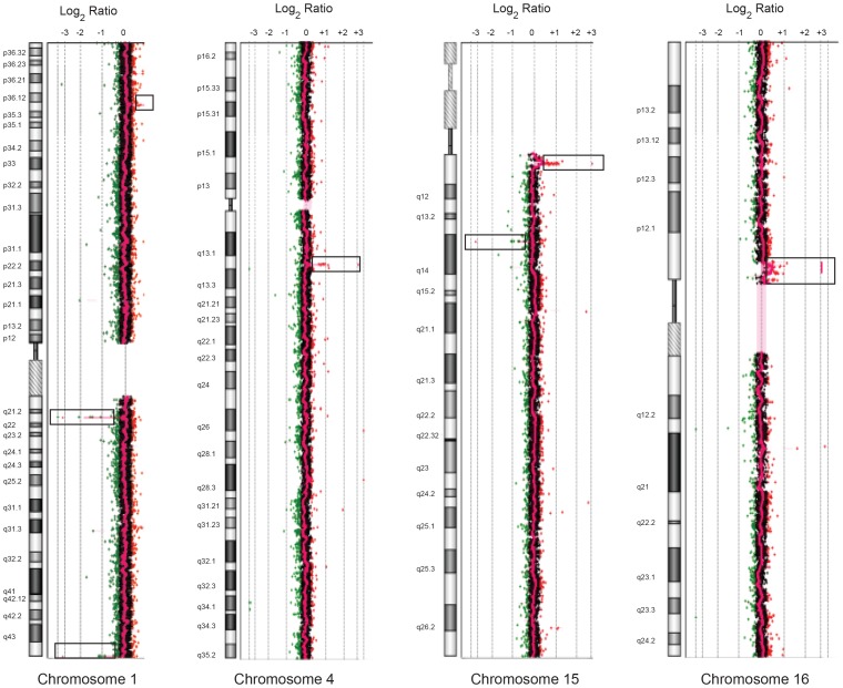
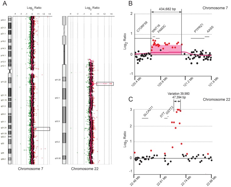
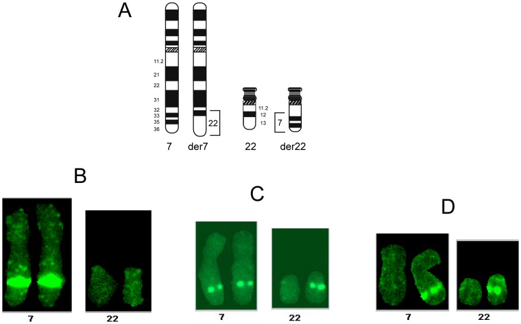
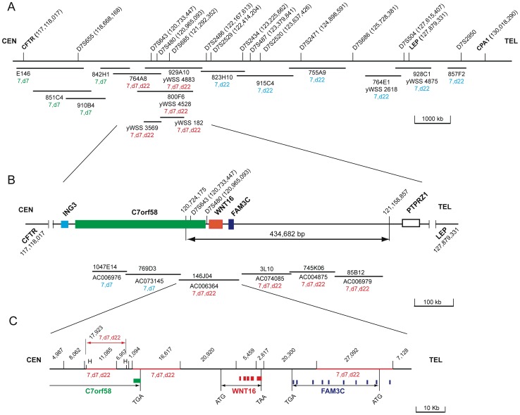
We characterized the t(7;22)(q32;q11.2) chromosomal translocation in an obese female with coarse features, short stature, developmental delay and a hypoplastic fifth digit. While these clinical features suggest Coffin-Siris Syndrome (CSS), we excluded a CSS diagnosis by exome sequencing based on the absence of deleterious mutations in six chromatin-remodeling genes recently shown to cause CSS. Thus, molecular characterization of her translocation could delineate genes that underlie other syndromes resembling CSS. Comparative genomic hybridization microarrays revealed on chromosome 7 the duplication of a 434,682 bp region that included the tail end of an uncharacterized gene termed C7orf58 (also called CPED1) and spanned the entire WNT16 and FAM3C genes. Because the translocation breakpoint on chromosome 22 did not disrupt any apparent gene, her disorder was deemed to result from the rearrangement on chromosome 7. Mapping of yeast and bacterial artificial chromosome clones by fluorescent in situ hybridization on chromosome spreads from this patient showed that the duplicated region and all three genes within it were located on both derivative chromosomes 7 and 22. Furthermore, DNA sequencing of exons and splice junctional regions from C7orf58, WNT16 and FAM3C revealed the presence of potential splice site and promoter mutations, thereby augmenting the detrimental effect of the duplicated genes. Hence, dysregulation and/or disruptions of C7orf58, WNT16 and FAM3C underlie the phenotype of this patient, serve as candidate genes for other individuals with similar clinical features and could provide insights into the physiological role of the novel gene C7orf58.
Conflict of interest statement
Figures
Similar articles
-
Clinical features of SMARCA2 duplication overlap with Coffin-Siris syndrome.Am J Med Genet A. 2016 Oct;170(10):2662-70. doi: 10.1002/ajmg.a.37778. Epub 2016 Jun 5. Am J Med Genet A. 2016. PMID: 27264538
-
SMARCE1, a rare cause of Coffin-Siris Syndrome: Clinical description of three additional cases.Am J Med Genet A. 2016 Aug;170(8):1967-73. doi: 10.1002/ajmg.a.37722. Epub 2016 Jun 5. Am J Med Genet A. 2016. PMID: 27264197 Free PMC article. Review.
-
Exome sequencing unravels unexpected differential diagnoses in individuals with the tentative diagnosis of Coffin-Siris and Nicolaides-Baraitser syndromes.Hum Genet. 2015 Jun;134(6):553-68. doi: 10.1007/s00439-015-1535-8. Epub 2015 Feb 28. Hum Genet. 2015. PMID: 25724810 Clinical Trial.
-
Coffin-Siris syndrome is a SWI/SNF complex disorder.Clin Genet. 2014 Jun;85(6):548-54. doi: 10.1111/cge.12225. Epub 2013 Jul 23. Clin Genet. 2014. PMID: 23815551
-
Coffin-Siris syndrome: phenotypic evolution of a novel SMARCA4 mutation.Am J Med Genet A. 2014 Jul;164A(7):1808-14. doi: 10.1002/ajmg.a.36533. Epub 2014 Apr 3. Am J Med Genet A. 2014. PMID: 24700502 Free PMC article. Review.
Cited by
-
Whole exome sequencing of a Saudi family and systems biology analysis identifies CPED1 as a putative causative gene to Celiac Disease.Saudi J Biol Sci. 2020 Jun;27(6):1494-1502. doi: 10.1016/j.sjbs.2020.04.011. Epub 2020 Apr 14. Saudi J Biol Sci. 2020. PMID: 32489286 Free PMC article.
-
Identification of IDUA and WNT16 Phosphorylation-Related Non-Synonymous Polymorphisms for Bone Mineral Density in Meta-Analyses of Genome-Wide Association Studies.J Bone Miner Res. 2016 Feb;31(2):358-68. doi: 10.1002/jbmr.2687. Epub 2015 Sep 11. J Bone Miner Res. 2016. PMID: 26256109 Free PMC article.
References
-
- Coffin GS, Siris E (1970) Mental retardation with absent fifth fingernail and terminal phalanx. Am J Dis Child 119: 433–439. - PubMed
-
- Tunnessen WW Jr, McMillan JA, Levin MB (1978) The Coffin-Siris syndrome. Am J Dis Child 132: 393–395. - PubMed
-
- Santen GW, Aten E, Sun Y, Almomani R, Gilissen C, et al. (2012) Mutations in SWI/SNF chromatin remodeling complex gene ARID1B cause Coffin-Siris syndrome. Nat Gen 44: 379–380. - PubMed
-
- Tsurusaki Y, Okamoto N, Ohashi H, Kosho T, Imai Y, et al. (2012) Mutations affecting components of the SWI/SNF complex cause Coffin-Siris syndrome. Nat Gen 44: 376–378. - PubMed
Publication types
MeSH terms
Substances
Supplementary concepts
Grants and funding
LinkOut - more resources
Full Text Sources
Medical
Miscellaneous
