. 2012;7(12):e53024.
doi: 10.1371/journal.pone.0053024. Epub 2012 Dec 31.

Robust formation and maintenance of continuous stratified cortical neuroepithelium by laminin-containing matrix in mouse ES cell culture

Affiliations

Robust formation and maintenance of continuous stratified cortical neuroepithelium by laminin-containing matrix in mouse ES cell culture

Makoto Nasu et al. PLoS One. 2012.

Abstract

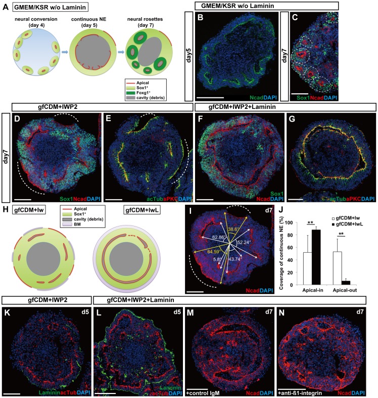
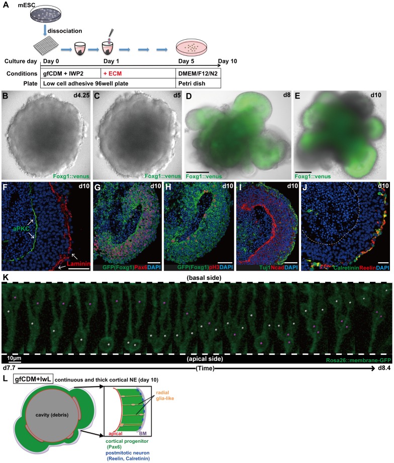
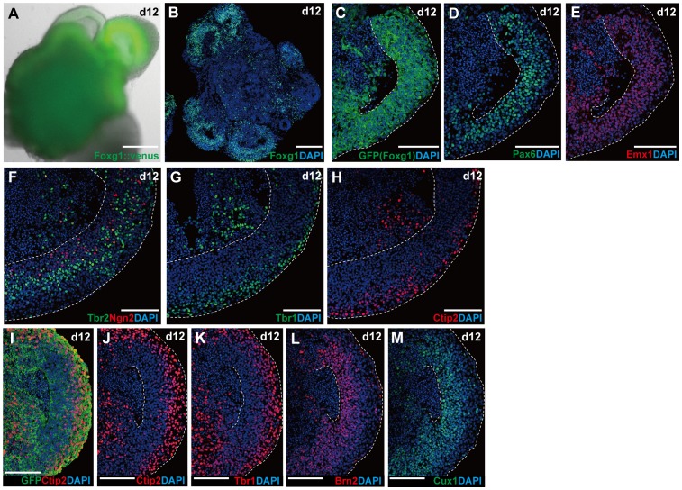
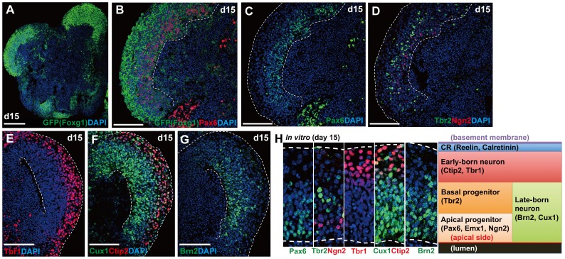
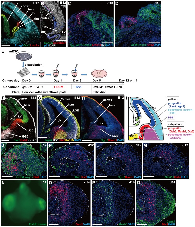
In the mammalian cortex, the dorsal telencephalon exhibits a characteristic stratified structure. We previously reported that three-dimensional (3D) culture of mouse ES cells (mESCs) can efficiently generate cortical neuroepithelium (NE) and layer-specific cortical neurons. However, the cortical NE generated in this mESC culture was structurally unstable and broke into small neural rosettes by culture day 7, suggesting that some factors for reinforcing the structural integrity were missing. Here we report substantial supporting effects of the extracellular matrix (ECM) protein laminin on the continuous formation of properly polarized cortical NE in floating aggregate culture of mESCs. The addition of purified laminin and entactin (a laminin-associated protein), even at low concentrations, stabilized the formation of continuous cortical NE as well as the maintenance of basement membrane and prevented rosette formation. Treatment with the neutralizing ß1-integrin antibody impaired the continuous NE formation. The stabilized cortical NE exhibited typical interkinetic nuclear migration of cortical progenitors, as seen in the embryonic cortex. The laminin-treated cortical NE maintained a continuous structure even on culture days 12 and 15, and contained ventricular, basal-progenitor, cortical-plate and Cajal-Retzius cell layers. The cortical NE in this culture was flanked by cortical hem-like tissue. Furthermore, when Shh was added, ventral telencephalic structures such as lateral ganglionic eminence-like tissue formed in the region adjacent to the cortical NE. Thus, our results indicate that laminin-entactin ECM promotes the formation of structurally stable telencephalic tissues in 3D ESC culture, and supports the morphogenetic recapitulation of cortical development.


Conflict of interest statement

Competing Interests: The authors have declared that no competing interests exist.

Figures

Figure 1
Figure 1. Efficient formation of continuous NE under chemically defined conditions.
(A) Schematic of the formation of neural rosettes under GMEM/KSR conditions. (B) Day-5 aggregate with partially continuous NE in culture with GMEM/KSR. (C) Day-7 aggregate with neural rosettes in culture with GMEM/KSR. Immunostaining for Sox1 and N-cadherin. A dashed circle indicates the unit of a neural rosette. (D–G) Effects of laminin ECM on the consistency of the A–B polarity in NE. Immunostaining of day-7 aggregates cultured in gfCDM+IWP2 (D–E) or gfCDM+IWP2+laminin ECM (F–G) for Sox1 and N-cadherin (D,F) or acetyl-α-tubulin and aPKC (E,G). (H) Schematic of SFEBq/gfCDM+Iw and SFEBq/gfCDM+IwL culture. (I–J) Quantification of consistency of A–B polarity in NE. (I) Schematic of quantification as a sum of angular degrees. The white or yellow arrows show the ends of each apical surface of apical-in or apical-out NE, respectively. A dashed line indicates an example of apical-out NE (D–E,I). (J) The percentage of continuous NE in gfCDM+IWP2 (white bars) and gfCDM+IWP2+laminin ECM (black bars) conditions. The values shown on graphs represent the mean ± s.e.m. **P<0.01. (K–L) The effect of laminin ECM on the formation/maintenance of laminin+ basement membrane (day 5). (K) The continuity of laminin+ basement membrane was lost in day-5 aggregate cultured without exogenous laminin ECM. (L) The day-5 aggregate cultured with laminin ECM was surrounded by continunous laminin+ basement membrane. (M–N) The effect of laminin ECM mediated by ß1-integrin. Anti-ß1-integrin impaired the formation of continuous NE in culture with laminin ECM. Note an increase of apical-out NE (N). No substantial effect was seen with control IgM (M). BM, basement membrane. Scale bars, 100 µm.
Figure 2
Figure 2. Formation of continuous cortical NE with a clear A–B polarity.
(A) Schematic of SFEBq/gfCDM+IwL culture. (B–C) Live imaging showing the formation of continuous NE in SFEBq/gfCDM+IwL culture on day 4.25 (B) and on day 5 (C). (D–E) Formation of continuous NE expressing Foxg1::venus on day 8 (D) and on day 10 (E). (F–J) Immunostaining of Foxg1::venus+ thick NE on day 10. (F) Immunostaining for the basement membrane marker laminin and the apical marker aPKC. (G) Cortical progenitors (Foxg1::venus+/Pax6+) occupied the majority of Foxg1::venus+ NE. (H) Mitotic cells (pH3+) located on the apical surface. (I) Postmitotic neurons (Tuj1+) in the superficial-most layer. (J) Immunostaining for Calretinin and Reelin. (K) Live imaging for interkinetic nuclear migration in continuous cortical NE generated from mESCs. Partial labeling was done by mixing Rosa26::membrane-GFP ESCs with non-labeled parental EB5 cells. Snapshots of two neural progenitors (days 7.7–8.4; also shown in Movie S2) undergoing interkinetic nuclear migrations (marked with white or pink dots). (L) Schematic of the formation of continuous cortical NE under gfCDM+IwL conditions on day 10. A dashed line indicates the apical border of NE. Scale bars, 200 µm (D–E); 50 µm (F–J); 10 µm (K).
Figure 3
Figure 3. Multi-layered cortical NE on day 12.
(A–B) Day-12 aggregates carrying continuous Foxg1::venus+ NE epithelia. (A) Fluorescent image of a Foxg1::venus+ aggregate. (B) Immunostaining for Foxg1. (C–H) Layered formation in continuous Foxg1::venus+ NE epithelia. (C–E) Cortical progenitors (Pax6+ and Emx1+) form a zone on the apical side of Foxg1::venus+ NE. (F) Immunostaining for Ngn2+ cells located in the basal part of the progenitor zone. A majority of basal progenitors (Tbr2+) were found outside of the Ngn2+ cell zone. (G–H) Early cortical plate neurons (Tbr1+, Ctip2+) were found on the basal side of Foxg1::venus+ NE epithelia. (I–M) Differential location of layer-specific cortical neurons in Foxg1::venus+ NE epithelia. (I–K) Immunostaining for Ctip2 (I–J) and Tbr1 (K). (L–M) Immunostaining for Brn2 (L) and Cux1 (M). Dashed lines indicate the apical and basal borders of NE. Scale bars, 200 µm (A–B); 100 µm (C–M).
Figure 4
Figure 4. Multi-layered cortical NE on day 15.
(A) Day-15 aggregate carrying continuous Foxg1::venus+ NE epithelia. Immunostaining for GFP. (B–G) Layer formation without the inside-out pattern. (B–C) Cortical progenitors (Foxg1::venus+/Pax6+) on the apical side. (D) Ngn2+ cells were located in a slightly deeper zone. Basal progenitors (Tbr2+) were outside of the Ngn2+ zone (E–G) Early cortical plate neurons (Tbr1+, Ctip2+) occupied the basal zone of continuous Foxg1::venus+ NE epithelia (E–F), while late cortical plate neurons (Brn2+, Cux1+) stayed on the apical side. (H) A schematic of cortical layer formation in vitro on day 15. Dashed lines indicate the apical or basal borders of NE. CR, Cajal-Retzius cell. Scale bars, 100 µm.
Figure 5
Figure 5. Formation and spatial arrangement of non-cortical telencephalic tissues.
(A) Expression pattern of markers for cortex, cortical hem, and choroid plexus on embryonic day 12 (E12). (B) Hem-derived Cajal-Retzius cells expressing p73 on E12. (C–D) The hem-like tissues (Lmx1a+, Otx2+) formed adjacent to cortical tissues (Foxg1::venus+) in culture. The hem-like tissues also contained p73+ cells. (E) Schematic for SFEBq/gfCDM+IwL culture with Shh treatment. (F–H) In vivo expression of pallial and subpallial markers on E12. Cortical markers (Pax6, Ngn2, Tbr1), LGE markers (Gsh2, Dlx2, Mash1, Gad65), and MGE markers (Nkx2.1, Dlx2, Mash1, Gad65). (I) Schematic of the marker expression pattern. (J–M) Shh treatment induced the formation of subpallial tissues. LGE tissues (Gsh2+, Dlx2+, Mash1+) were located between cortical tissues (Pax6+, Ngn2+, Tbr1+) and MGE tissues (Nkx2.1+, Dlx2+, Mash1+). Arrowheads indicate a transition between pallial and subpallial tissues. (N) Expression of Gsh2::venus in thickened NE tissue on day 14. (O–Q) Immunostaing of Gsh2::venus+ NE on day 14. Immunostaining for Gsh2 (O), Mash1 (P) and Gad67/Dlx2 (Q). chp, choroid plexus; LGE, lateral ganglionic eminence; LV, lateral ventricle; MGE, medial ganglionic eminence; PSB, pallial-subpallial boundary; Th, thalamus. Scale bars, 100 µm (A–D,J–M,O–Q); 200 µm (F–H,N).

References

    1. Rallu M, Corbin JG, Fishell G (2002) Parsing the prosencephalon. Nature Rev Neurosci 3: 943–951. - PubMed
    1. Shimamura K, Hartigan DJ, Martinez S, Puelles L, Rubenstein JL (1995) Longitudinal organization of the anterior neural plate and neural tube. Development 121: 3923–3933. - PubMed
    1. Stoykova A, Treichel D, Hallonet M, Gruss P (2000) Pax6 modulates the dorsoventral patterning of the mammalian telencephalon. J Neurosci 20: 8042–8050. - PMC - PubMed
    1. Hevner RF, Daza RAM, Rubenstein JLR, Stunnenberg H, Olavarria JF, et al. (2003) Beyond laminar fate: toward a molecular classification of cortical projection/pyramidal neurons. Dev Neurosci 25: 139–151. - PubMed
    1. Shen Q, Wang Y, Dimos JT, Fasano CA, Phoenix TN, et al. (2006) The timing of cortical neurogenesis is encoded within lineages of individual progenitor cells. Nature neurosci 9: 743–751. - PubMed

Publication types