. 2012;7(12):e53098.
doi: 10.1371/journal.pone.0053098. Epub 2012 Dec 31.

Defective soil for a fertile seed? Altered endometrial development is detrimental to pregnancy success

Affiliations

Defective soil for a fertile seed? Altered endometrial development is detrimental to pregnancy success

Jemma Evans et al. PLoS One. 2012.

Abstract

Background: Synchronous development of the endometrium (to achieve a receptive state) and of the embryo is essential for successful implantation and ongoing pregnancy. Endometrial receptivity exists only for a finite time in a menstrual cycle and the endometrium is refractory to embryo implantation outside of this window. Administration of hormones to stimulate multifollicular development within the ovary, integral to the majority of assisted reproduction (ART) protocols, dramatically alters the hormonal milieu to which the endometrium is exposed versus normal menstrual cycles. Endometrial maturation may be profoundly affected by this altered endocrine environment.

Aim: Compare endometrial histology in fertile women, fertile women undergoing hormonal stimulation for oocyte donation and infertile women undergoing fresh embryo transfers in an ART cycle with further comparisons between women who did or did not become pregnant. Examine the presence of leukocytes and markers of endometrial maturation.

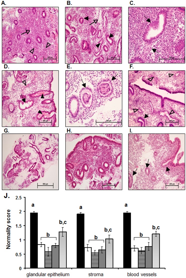
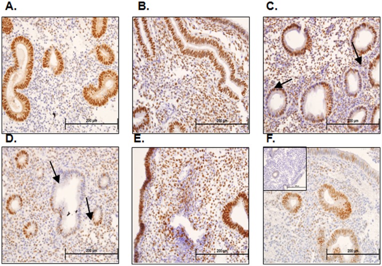
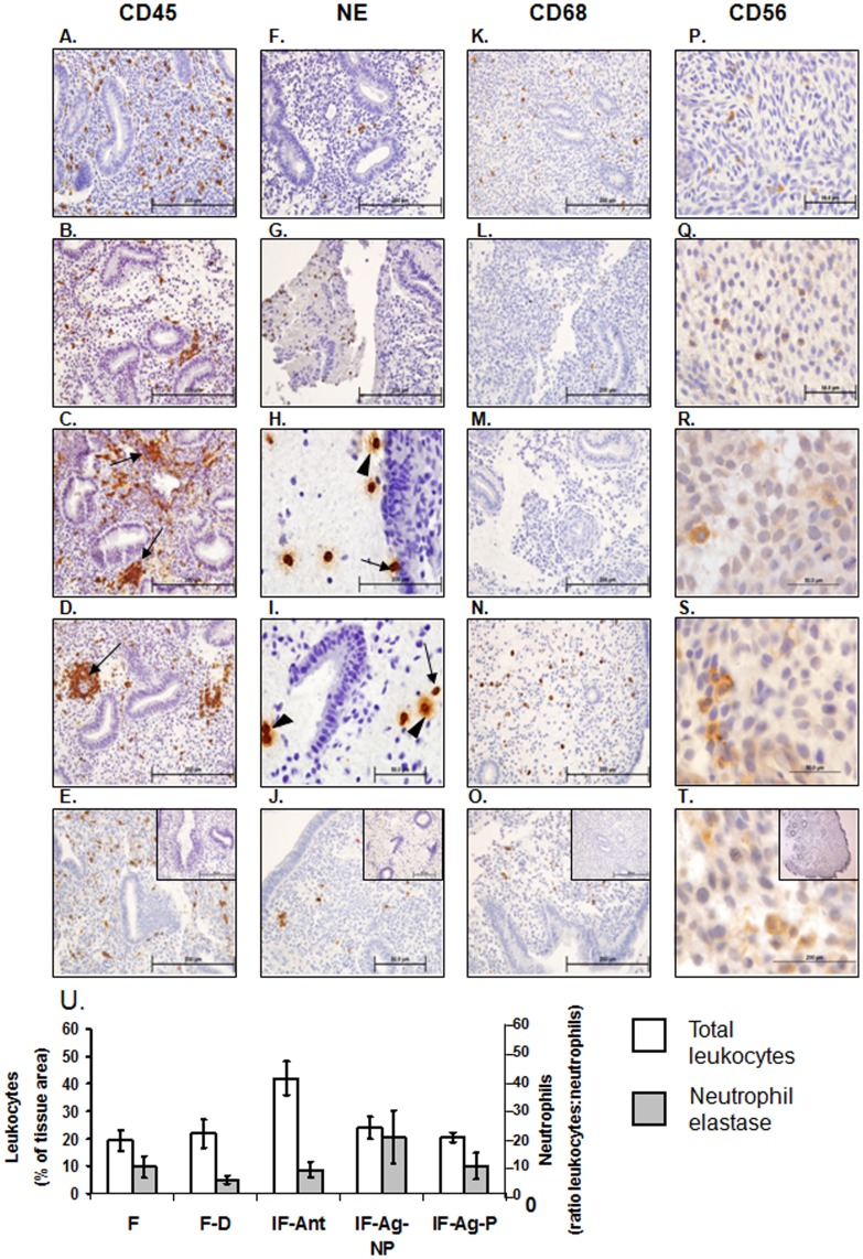
Methods: Endometrial histology was examined by hematoxylin and eosin staining with a semi quantitative scoring method developed to compare histological appearance of tissues. The presence of leukocytes and developmental markers was examined by immunohistochemistry and scored.

Results: Endometrial histology was dramatically altered upon stimulation for ART. However, those women who became pregnant presented with significantly less alterations in histological endometrial maturation. Numbers and activation status of leukocyte populations were also altered within the endometria stimulated for ART, with neutrophils undergoing degranulation, usually observed only pre-menstrually.

Conclusion: We propose that such developmental changes render the endometrium hostile to the embryo and that modifications to ART protocols should be considered to take account of the requirement for endometrial receptivity and hence increase pregnancy rates.


Conflict of interest statement

Competing Interests: The authors have read the journal's policy and have the following conflicts: J.E, N.J.H., and C.H. have nothing to declare. L.R. and L.A.S. have received research grants for this research. The other authors have the following interests. Monash IVF, though its research foundation, contributed funding for the study and L.R is a shareholder and company board member. There are no patents, products in development or marketed products to declare. This does not alter the authors' adherence to all PLOS ONE policies on sharing data and materials as detailed in the online guide for authors.

Figures

Figure 1
Figure 1. Endometrial histology of IVF stimulated and non-stimulated endometria.
In the fertile non-stimulated endometrium at LH+2 (A) the glands are small and straight with only very early signs of secretory changes. The stroma is compact with minor signs of oedema (open arrow), and blood vessels are small (open arrow head). At hCG+2 in the fertile donor endometrium stimulated with GnRH agonist, (B & C) the glandular epithelial cells show secretory changes as evidenced by sub-nuclear vacuoles (C, closed arrows), some evidence of secretory activity can also be observed within the lumen of some endometrial glands (B, closed arrowhead). The stroma is expanded and oedematous (B, open arrow). At hCG+2 in the infertile endometrium stimulated with the GnRH antagonist protocol (D & E) a range of different endometrial cycle stage appearances presented. In some endometria (D) the endometrial glandular epithelial cells have lost their sub-nuclear vacuoles and are highly secretory (D, closed arrowheads) with glands becoming tortuous (D, arrow). The stroma is also highly oedematous and large open blood vessels are present (D, open arrowhead). Other infertile GnRH antagonist stimulated endometria (E) present with sub-nuclear vacuoles evident in the endometrial glands (E, arrows) and expanded stroma. In the endometria at hCG+2 of infertile women stimulated with the GnRH agonist protocol who did not subsequently become pregnant (F & G) a mixed picture of endometrial histology is observed. Sub-nuclear vacuoles are present in the endometrial glandular epithelial cells (F, closed arrow) with the presence of expanded oedematous stroma and large expanded blood vessels close to the luminal epithelium (F, open arrowheads). In other endometria from this group (G) the tissue histology is disturbed with highly developed, tortuous, secretory endometrial glands and highly oedematous stroma, with the tissue as a whole presenting a fragile appearance. In infertile women stimulated with the GnRH agonist protocol who subsequently became pregnant (H & I) fairly small compact endometrial glands with only early signs of secretory transformation (I, arrows) were observed at hCG+2. The stroma, while expanded in some areas is generally compact and the blood vessels are not highly developed. Endometrial histology was scored by means of a ‘normality score’ (J) with tissues of normal histology for LH+2 allocated a score of 2, tissues with somewhat changed histology allocated a score of 1 and tissues with highly altered histology allocated a score of 0. Data for fertile women are presented in black (?), data for fertile donors are presented in white (□), data for infertile GnRH antagonist are presented in horizontal lines (≡), data for infertile GnRH agonist not pregnant are presented in dark grey (?), data for infertile GnRH agonist pregnant are presented in light grey (?). Data are presented as mean ± SEM. a is significantly different from b, p<0.01; b is significantly different from c, p<0.05.
Figure 2
Figure 2. Progesterone receptor immunohistochemistry and semi-quantitative analysis.
Progesterone receptor (PR) consistently immunolocalizes to the glandular epithelial cells of endometria taken from fertile women at LH+2 (A). In fertile ovum donors stimulated with the GnRH protocol PR immunostaining is localized to glandular epithelial cells, luminal epithelial cells and stromal cells on hCG+2 (B). PR staining is similarly patchy in the glandular epithelial cells of endometria taken from infertile women stimulated with GnRH antagonist at hCG+2 (C & D, arrows) and in the stroma (C & D). PR immunostaining localizes to the glandular epithelial cells, luminal epithelial cells and stromal cells of endometria of infertile women undergoing the GnRH agonist protocol who did not subsequently become pregnant at hCG+2 (E). PR immunolocalized mainly to the glandular epithelium with patchy luminal epithelial and stromal staining in endometria of infertile women undergoing the GnRH agonist protocol who subsequently become pregnant at hCG+2 (F).
Figure 3
Figure 3. Leukocyte immunohistochemistry and semi quantitative analysis.
Immunohistochemical staining was performed to examine the localization leukocyte subtypes (CD45/leukocyte common antigen, A-E), neutrophils (neutrophil elastase, F-J), macrophages (CD68, K-O) and uterine natural killer cells (CD56, P-R). In fertile endometria at LH+2 leukocytes are scattered throughout the tissue (A). At hCG+2 in stimulated endometria from fertile donor women undergoing GnRH agonist stimulation (B), infertile women undergoing GnRH antagonist stimulation (C) and infertile women undergoing GnRH agonist stimulation who did not become pregnant (D) clusters of leukocytes can be observed particularly adjacent to endometrial glands and surrounding blood vessels (B, C, and D, arrows). In endometria of women stimulated with the GnRH agonist protocol who subsequently became pregnant at hCG+2 (E) leukocytes are found scattered throughout the tissue, similar to that observed in fertile women at LH+2 (A). Very few neutrophils can be observed in the endometria of fertile women at LH+2 (F) or fertile women undergoing GnRH agonist stimulation for ovum donation at hCG+2 (G). In stimulated endometria from GnRH antagonist stimulated infertile women (H) or GnRH agonist protocol who did not become pregnant (I) numerous neutrophils can be observed particularly within mucous like areas and to a lesser degree within the tissue. Many of these are degranulating (H & I, closed arrowhead). Very few neutrophils were evident in the endometria of GnRH agonist stimulated infertile women who became pregnant (J). Scattered macrophages could be observed in endometria of fertile women at LH+2 (K). Very few macrophages were present in fertile women undergoing GnRH agonist stimulation for ovum donation (L) or infertile women undergoing GnRH antagonist stimulation (M). Scattered macrophages were observed in the endometria of infertile women stimulated with the GnRH agonist protocol whether they did not (N) or subsequently achieved (O) pregnancy. Few uterine natural killer cells were detected at LH+2 (P) or hCG+2 in endometria of fertile (Q) infertile women stimulated with the GnRH antagonist protocol (R), or the GnRH agonist protocol who did not (S) or did (T) become pregnant at hCG+2. IgG controls are shown inset in panels E, J, O and T. Scale bars are shown in each image, all 200 µm except P - T at 50 µm. Semi quantitative scoring of total leukocyte numbers (U) revealed increased leukocyte numbers in endometria of infertile women stimulated with the GnRH antagonist protocol at hCG+2 compared with fertile women at LH+2 (U, white bars, P<0.05). No significant differences in neutrophil numbers were observed (U, grey bars). F = fertile, IF = infertile, F–D = fertile donor, IF-Ant = infertile antagonist, IF-Ag-NP = infertile agonist non-pregnant, IF-Ag-P = infertile agonist pregnant. Data are expressed as mean ± SEM, *P<0.05.
Figure 4
Figure 4. Vasculature/CD34 and decidualization/prolactin immunostaining.
Blood vessels within the endometria of fertile women at LH+2 were generally small and compact (A). Examination of endometria from fertile women stimulated with the GnRH agonist protocol for ovum donation at hCG+2 revealed a mixture of small blood vessels and grossly enlarged blood vessels (B, arrow). Endometria from infertile women stimulated with the GnRH antagonist protocol at hCG+2 revealed a mixed picture of vascular development, with some endometria presenting grossly enlarged blood vessels (C, arrow) whereas some presented with small compact blood vessels (D). Women stimulated with the GnRH agonist protocol at hCG+2 who did not subsequently become pregnant presented with large endometrial blood vessels (E), whereas women stimulated with this protocol who did become pregnant generally presented with smaller more compact blood vessels (F). Fertile women at LH+2 had little to no immunostaining for prolactin within the endometrial stromal compartment (G). Some areas within the endometria of fertile women stimulated with the GnRH protocol for ovum donation (H), infertile women stimulated with the GnRH antagonist protocol (I) and infertile women stimulated with the GnRH agonist protocol who did not become pregnant (J) at hCG+2 demonstrated intense stromal prolactin immunostaining indicative of decidual changes. Endometria from infertile women stimulated with the GnRH agonist protocol at hCG+2 who subsequently became pregnant (K) demonstrated little stromal prolactin immunostaining, but did demonstrate glandular prolactin immunostaining indicative of secretory changes (K, arrows).

References

    1. Salamonsen LA, Nie G, Hannan NJ, Dimitriadis E (2009) Society for Reproductive Biology Founders' Lecture 2009. Preparing fertile soil: the importance of endometrial receptivity. Reprod Fertil Dev 21: 923–934. - PubMed
    1. van der Gaast MH, Classen-Linke I, Krusche CA, Beier-Hellwig K, Fauser BC, et al. (2008) Impact of ovarian stimulation on mid-luteal endometrial tissue and secretion markers of receptivity. Reprod Biomed Online 17: 553–563. - PubMed
    1. Boomsma CM, Kavelaars A, Eijkemans MJ, Amarouchi K, Teklenburg G, et al. (2009) Cytokine profiling in endometrial secretions: a non-invasive window on endometrial receptivity. Reprod Biomed Online 18: 85–94. - PubMed
    1. Boomsma CM, Kavelaars A, Eijkemans MJ, Lentjes EG, Fauser BC, et al. (2009) Endometrial secretion analysis identifies a cytokine profile predictive of pregnancy in IVF. Hum Reprod 24: 1427–1435. - PubMed
    1. Boomsma CM, Kavelaars A, Eijkemans MJ, Fauser BC, Heijnen CJ, et al. (2010) Ovarian stimulation for in vitro fertilization alters the intrauterine cytokine, chemokine, and growth factor milieu encountered by the embryo. Fertil Steril 94: 1764–1768. - PubMed

Publication types